
Extended Total Maxillectomy with Infratemporal Fossa, Transpterygoid and Anterior Skull Base Clearance for Sinonasal Mucormycosis
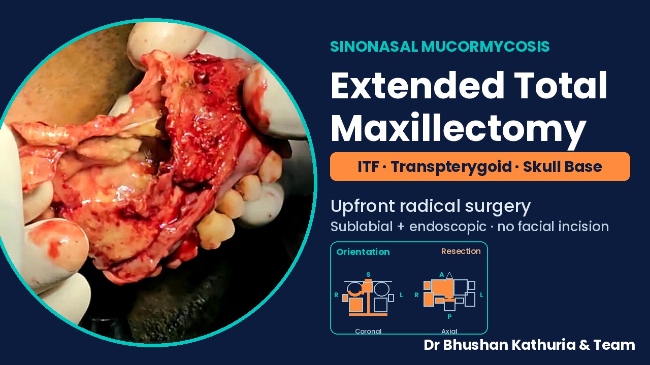
Upfront radical resection for fulminant sinonasal mucormycosis. Intended audience: ENT residents, head and neck and skull base surgeons.
- Diagnosis
- Fulminant sinonasal mucormycosis
- Procedure
- Extended total maxillectomy with ITF, transpterygoid and anterior skull base clearance
- Approach
- Hybrid sublabial + endoscopic; no facial incision
- Rehabilitation
- Obturator / prosthesis
- Video
- 4 min · step-by-step
Operative video with step captions and geometric orientation markings. Summary video.
Abstract
Fulminant sinonasal mucormycosis is an angioinvasive fungal infection in which the visible eschar represents only part of the devitalised tissue. Drawing on a surgical experience of more than 300 cases in the post-COVID surge, we describe an upfront radical, single-stage resection: extended total maxillectomy with infratemporal fossa (ITF), transpterygoid and anterior skull base clearance, performed through a hybrid sublabial and endoscopic approach without a facial incision. The operative sequence, the extent of resection with geometric orientation, and the endoscopic review of the surgical bed are presented step by step with the full operative video.
Keywords: mucormycosis; rhino-orbital mucormycosis; extended total maxillectomy; infratemporal fossa; transpterygoid approach; anterior skull base; endoscopic sinus surgery; sublabial approach
Introduction
Rhino-orbito-cerebral mucormycosis rose sharply in India during and after the COVID-19 pandemic, predominantly in patients with uncontrolled diabetes and recent corticosteroid exposure. Mucorales invade vessel walls, causing thrombosis and infarction of the surrounding tissue. Systemic antifungals reach only perfused tissue; surgical removal of all non-viable tissue is therefore central to survival [1,2].
Surgical philosophy varies between limited, staged endoscopic debridement and upfront radical resection. In fulminant disease involving the maxilla, palate, pterygopalatine and infratemporal fossae and the ethmoid–sphenoid complex, our approach is a single-stage radical resection to bleeding, viable margins in all directions, accepting a defect that is later rehabilitated with an obturator or prosthesis.
Case presentation
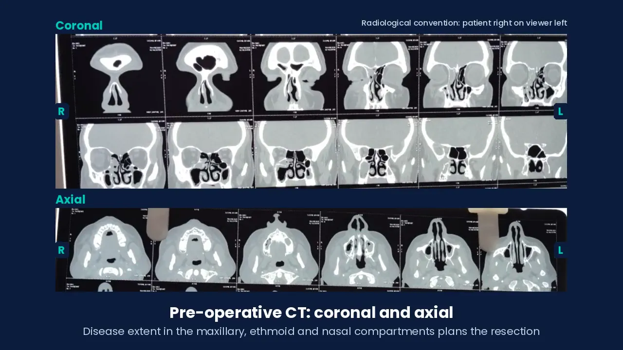
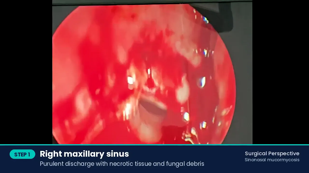
A patient with fulminant sinonasal mucormycosis is presented. Typical presenting features in our series include uncontrolled diabetes or recent corticosteroid use with nasal obstruction, blackish nasal discharge, facial numbness and loosening of teeth. In this patient, diagnostic endoscopy showed fungal debris and necrotic tissue in the frontal recess and purulent discharge with necrotic tissue in the right maxillary sinus. Pre-operative CT (coronal and axial) defined the extent of maxillary, ethmoid and nasal disease and planned the resection.
Management followed our protocol of urgent surgery alongside antifungal therapy and metabolic correction. Written informed consent for surgery and for de-identified educational use of operative images was obtained.
Surgical technique
Diagnostic endoscopy
With a 70° endoscope, fungal debris and necrotic tissue are seen in the frontal recess; the right maxillary sinus contains purulent discharge with necrotic tissue. Transoral endoscopy assesses the palatal and alveolar mucosa.
Diagnostic endoscopy Sublabial (gingivobuccal) incision
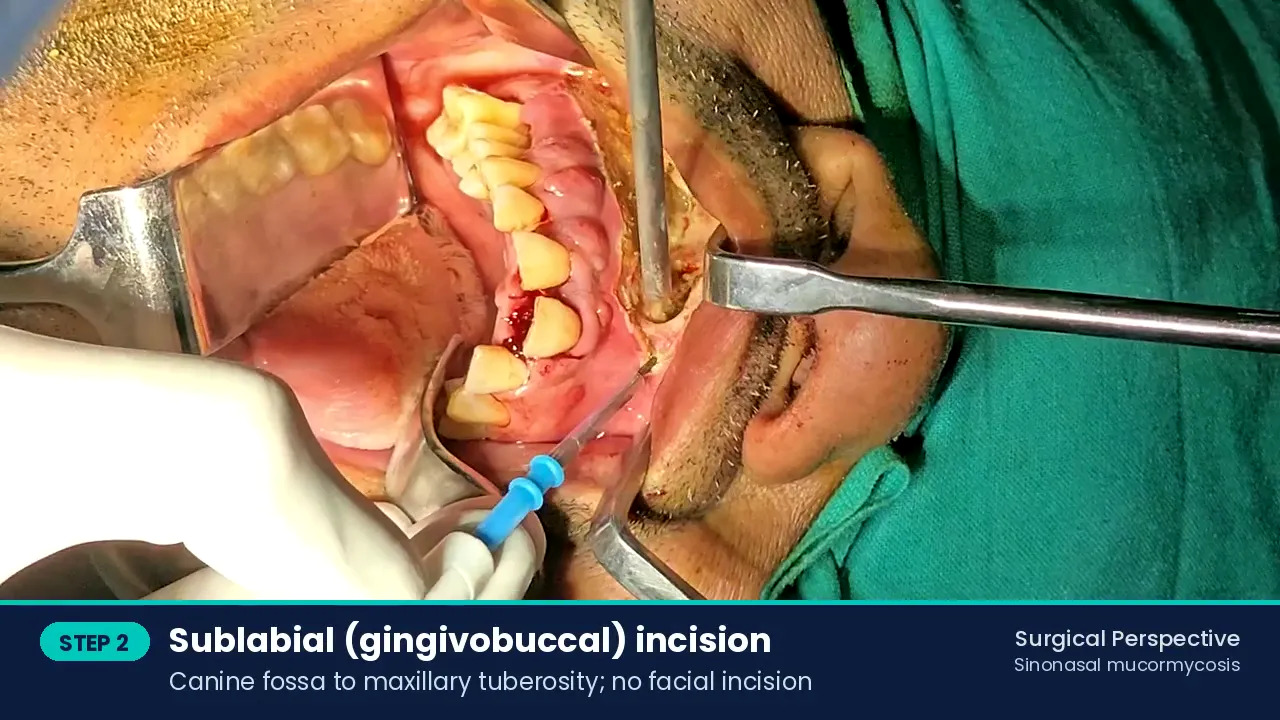
Incision from the canine fossa to the maxillary tuberosity; no facial incision is used.
Sublabial (gingivobuccal) incision Subperiosteal exposure
The anterior wall of the maxilla, zygomatic buttress and infraorbital rim are exposed in the subperiosteal plane.
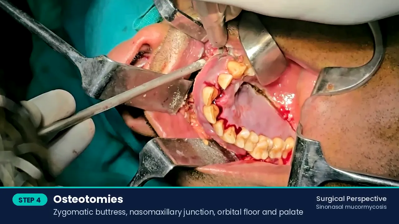
Osteotomies
Bone cuts with a powered drill through the zygomatic buttress, nasomaxillary junction, orbital floor and palate.
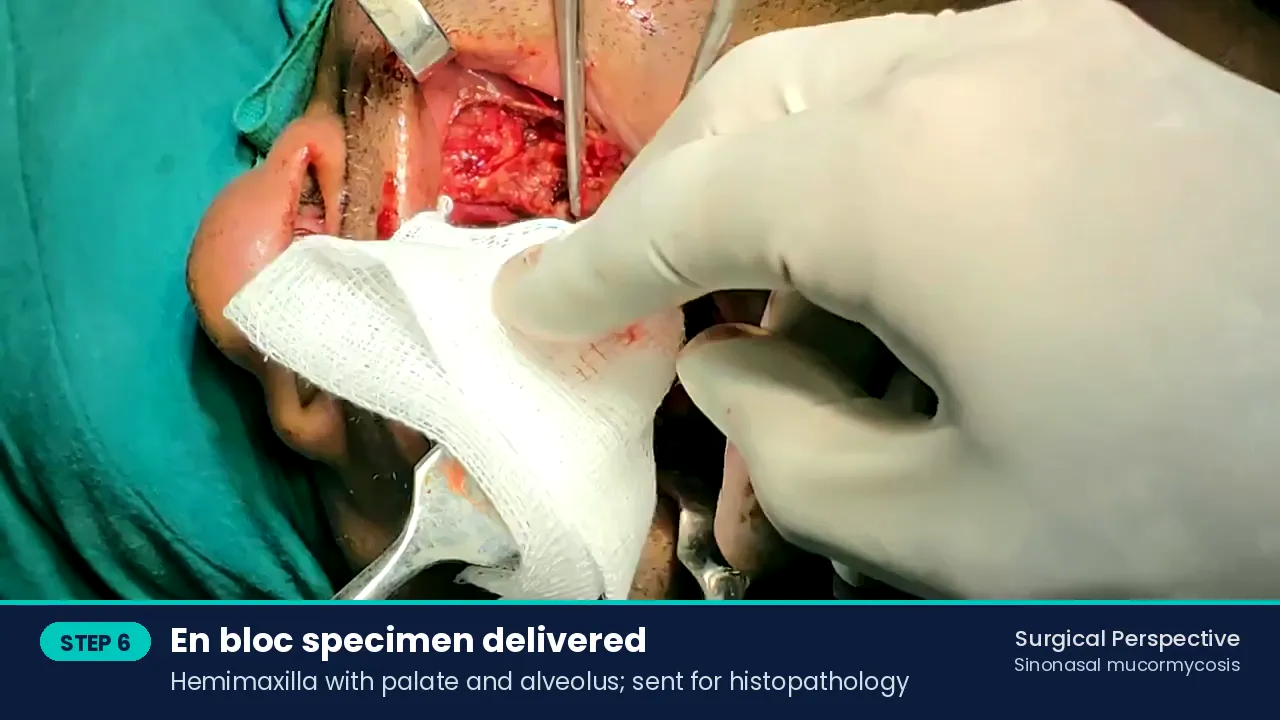
Osteotomies Extended total maxillectomy and en bloc delivery
The maxilla is mobilised with the palate, alveolus and pterygoid attachments and delivered en bloc for histopathology.
Extended total maxillectomy and en bloc delivery Debridement of the ITF and pterygoid region
Necrotic soft tissue is cleared to bleeding, viable margins (transpterygoid).
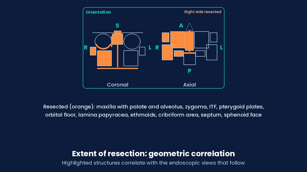
Endoscopic review of the surgical bed
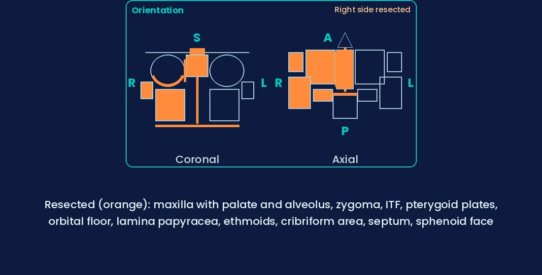
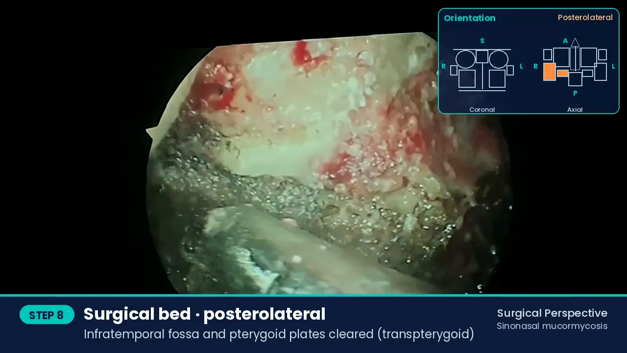
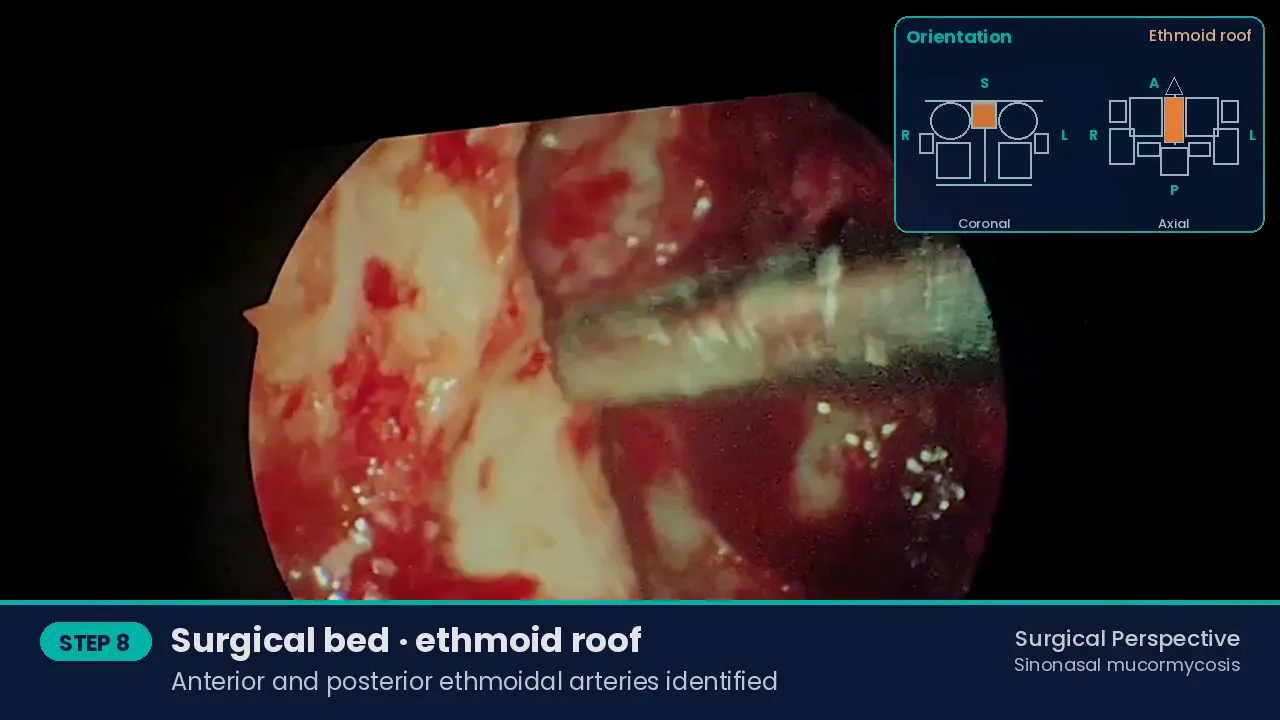
Lateral to medial and anterior to posterior: drilled zygoma laterally; ITF and pterygoid plates; orbital floor removed anteriorly with the lamina papyracea in view; anterior and posterior ethmoidal arteries; cribriform area and planum; anterior wall of the sphenoid removed; septum resected medially.
Endoscopic review of the surgical bed Final inspection and packing
Haemostasis is confirmed and the cavity is packed in preparation for the obturator.
Postoperative management
- Liposomal amphotericin B continued with step-down to posaconazole or isavuconazole, strict glycaemic control and correction of ketoacidosis.
- Serial endoscopic cavity review; early re-debridement of any fresh necrosis.
- Interim obturator, followed by a definitive prosthesis for speech, mastication and contour.
Discussion
Upfront radical surgery is mutilating, but in fulminant angioinvasive disease it is life-saving: it removes the fungal load and the non-perfused tissue that antifungals cannot reach. The hybrid route — sublabial for the maxilla, palate and ITF; endoscopic for the ethmoid, sphenoid and skull base — gives wide access without facial scars.
Surgery, antifungal therapy and metabolic control act only together. The resulting defect is accepted in favour of survival and is rehabilitated prosthetically.
Learning points
- Mucormycosis is the tip of the iceberg: resect beyond the visible eschar to bleeding, viable margins.
- Single-stage radical resection rather than serial limited debridement in fulminant disease.
- Hybrid sublabial + endoscopic access avoids a facial incision.
- Rehabilitate the defect with an obturator or prosthesis.
References
- Cornely OA, Alastruey-Izquierdo A, Arenz D, et al. Global guideline for the diagnosis and management of mucormycosis: an initiative of the ECMM in cooperation with the Mycoses Study Group Education and Research Consortium. Lancet Infect Dis. 2019;19(12):e405-e421. https://doi.org/10.1016/S1473-3099(19)30312-3
- Honavar SG. Code Mucor: guidelines for the diagnosis, staging and management of rhino-orbito-cerebral mucormycosis in the setting of COVID-19. Indian J Ophthalmol. 2021;69(6):1361-1365. https://doi.org/10.4103/ijo.IJO_1165_21
Consent and ethics
Written informed consent was obtained for surgery and for de-identified use of clinical images and video for education. No identifying information is shown; eyes are blurred where visible. This report is for medical education and does not replace individual clinical advice.
Surgical team
Dr Bhushan Kathuria & Team, Santosh Advance ENT Healthcare, Hisar. Consultants: Dr Bhushan Kathuria, Dr Parmod Jangra, Dr Harshvardhan. Clinical fellows: Dr Ayush, Dr Iqrorjon. Team · Fellowship and previous fellows.
How to cite
Kathuria B, and Team. Extended Total Maxillectomy with Infratemporal Fossa, Transpterygoid and Anterior Skull Base Clearance for Sinonasal Mucormycosis. Surgical Perspective · Surgical Case Reports. 2026. Available at: https://bhushankathuria.com/case-sinonasal-mucormycosis-extended-total-maxillectomy.html
More case reports
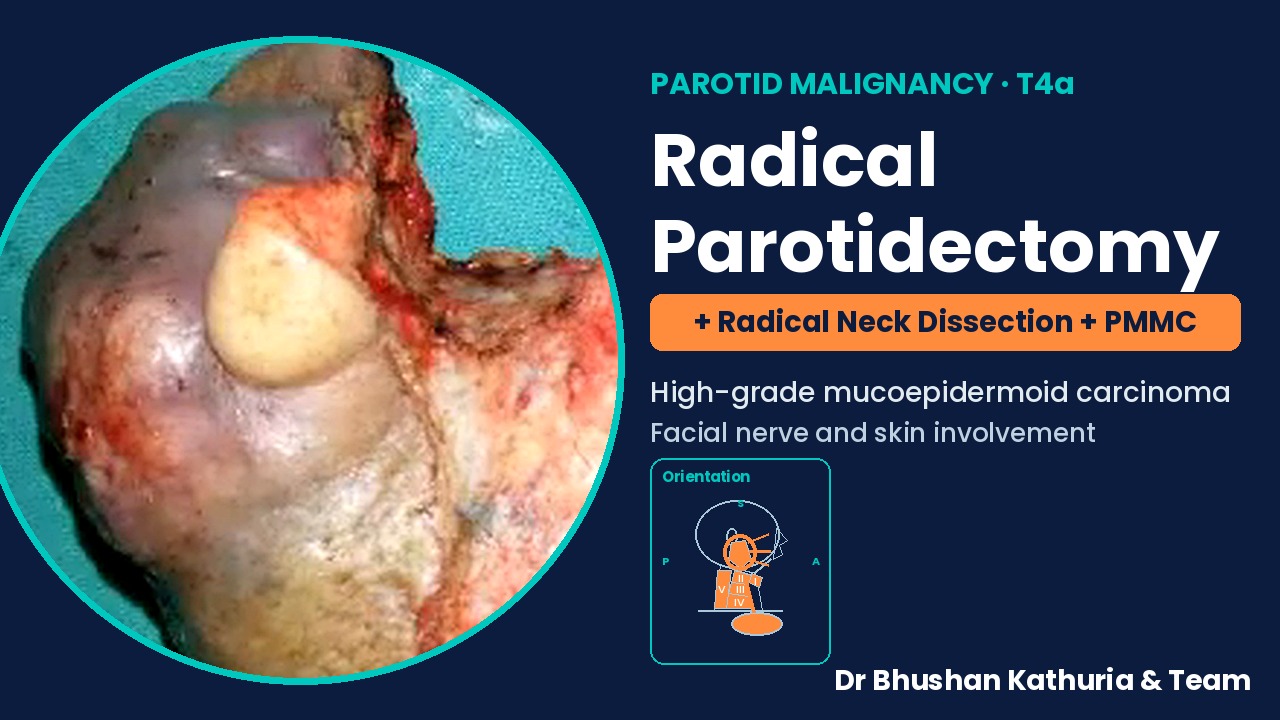
High-Grade Mucoepidermoid Carcinoma of the Parotid with Facial Nerve and Skin Involvement: Radical Parotidectomy, Radical Neck Dissection and PMMC Flap Reconstruction
Composite parotid resection with single-stage PMMC reconstruction
▶ Video + case report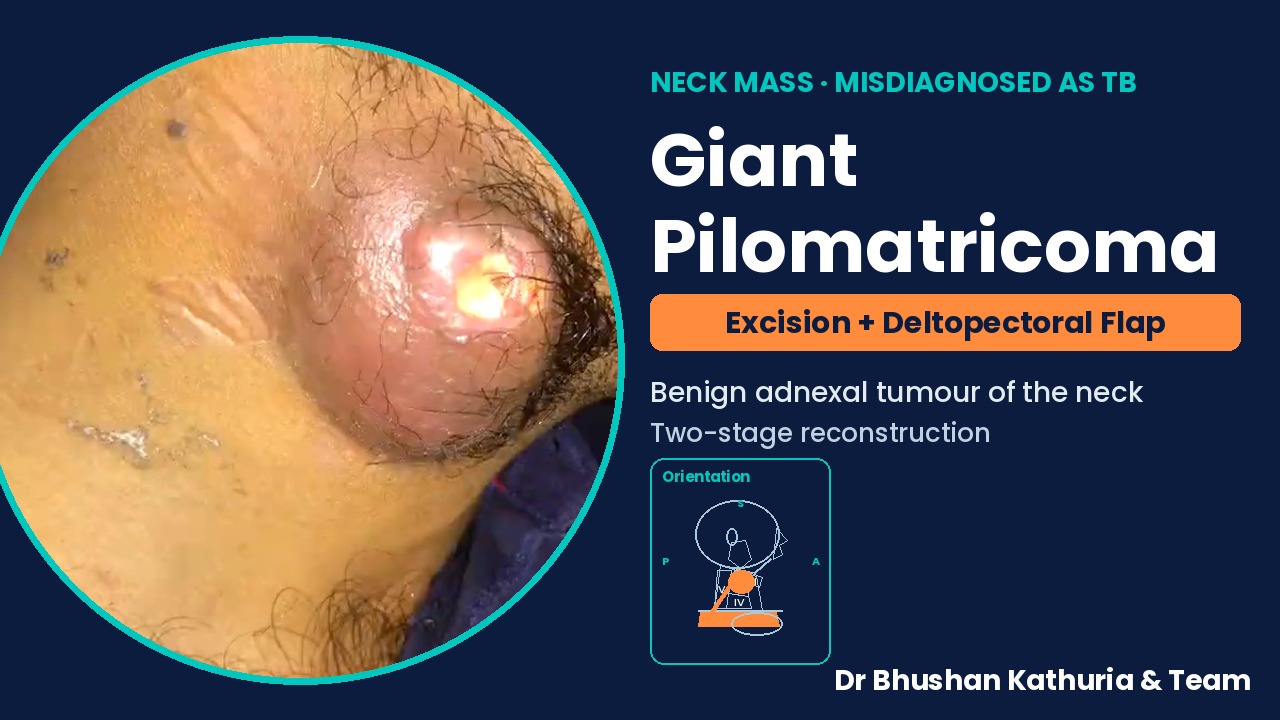
Giant Pilomatricoma of the Neck Misdiagnosed as Tubercular Lymphadenitis: Wide Local Excision and Two-Stage Deltopectoral Flap Reconstruction
Pilomatricoma presenting as a non-healing neck mass
▶ Video + case report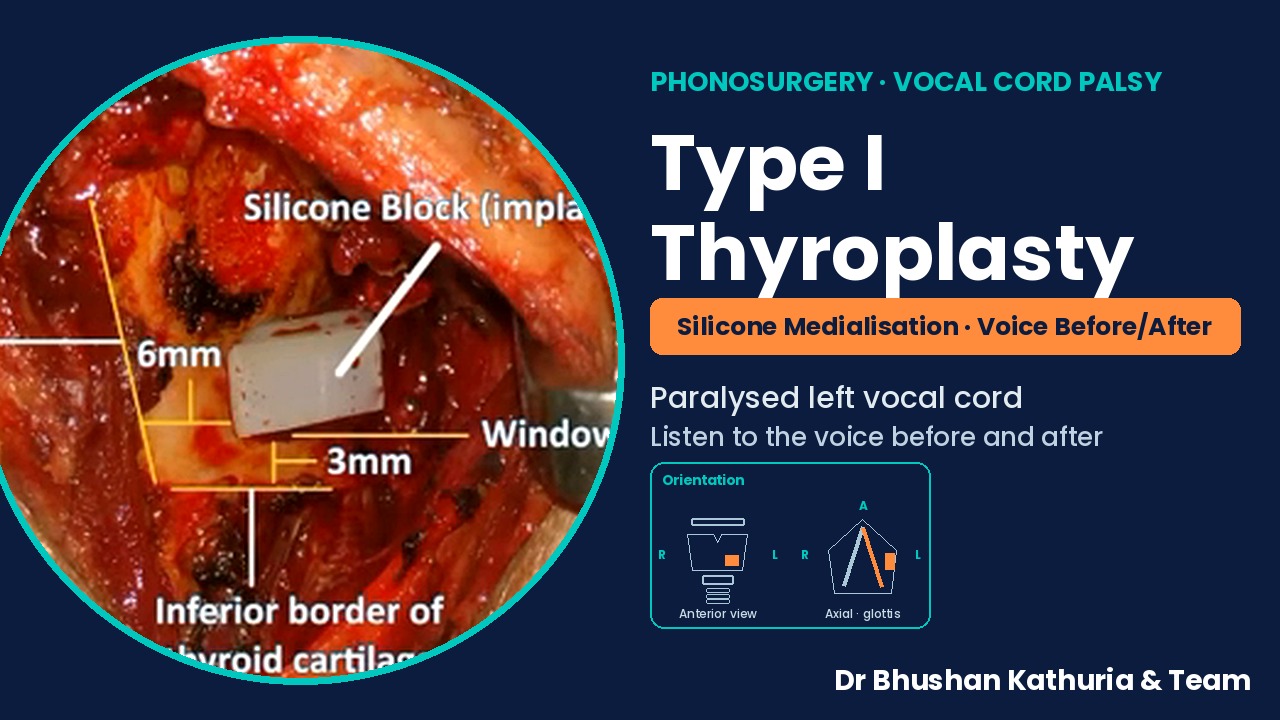
Type I Thyroplasty: Silicone Medialisation of a Paralysed Left Vocal Cord, with Pre- and Post-operative Voice
Medialisation thyroplasty for unilateral vocal fold palsy
▶ Video + case report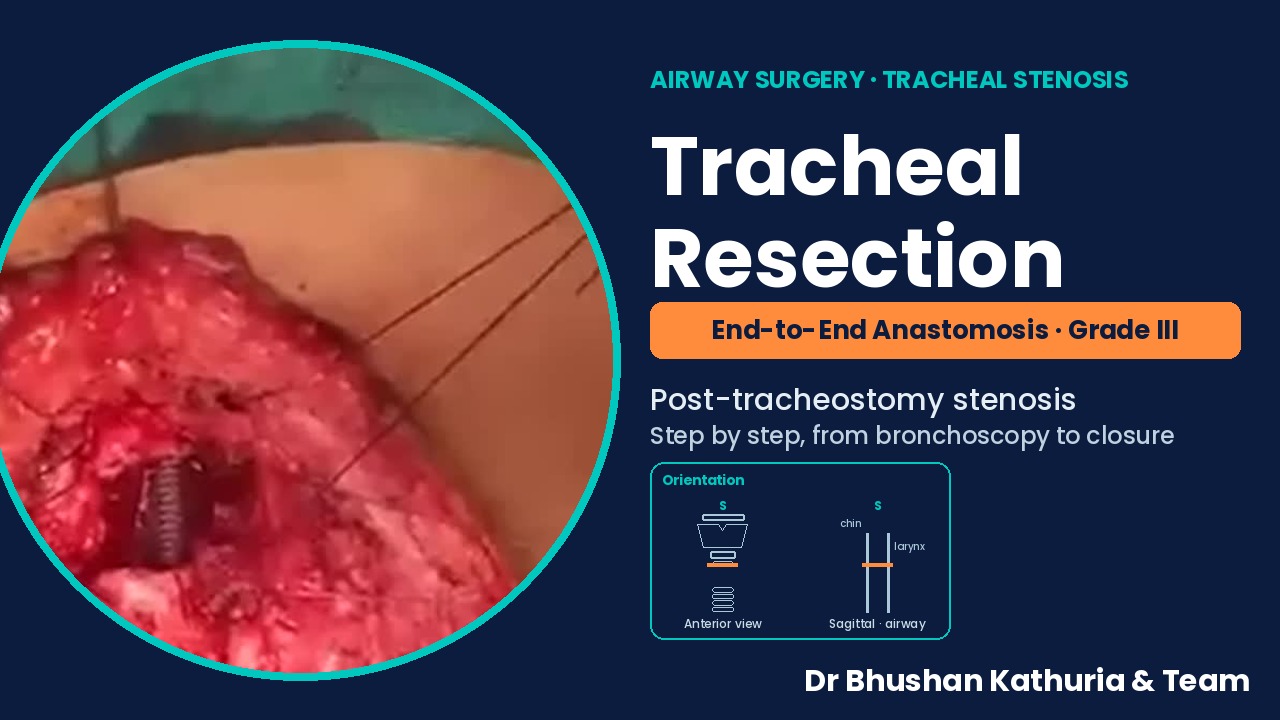
Segmental Tracheal Resection and End-to-End Anastomosis for Grade III Post-tracheostomy Tracheal Stenosis
Tracheal resection and primary anastomosis after failed decannulation
▶ Video + case report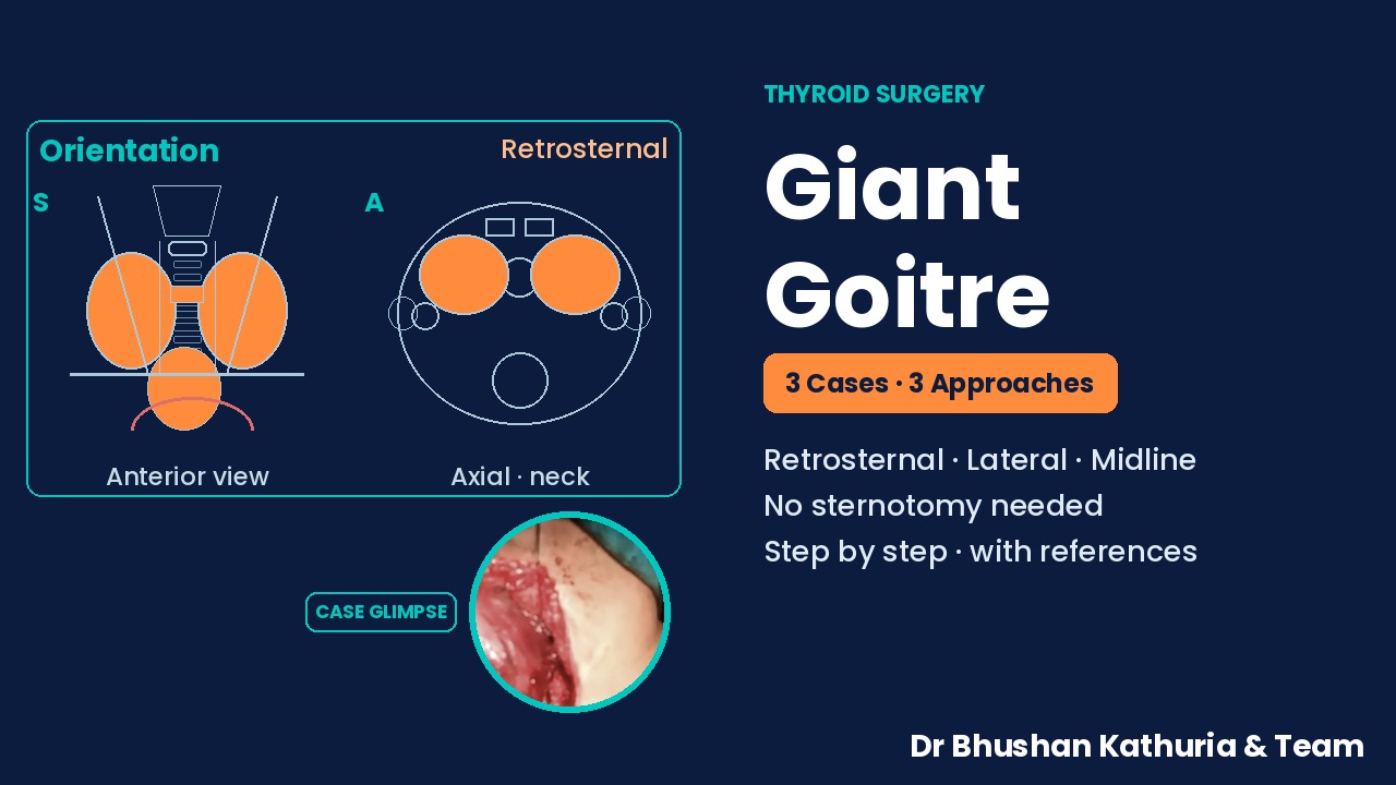
Giant Goitre: Three Cases and Three Approaches — Cervical Delivery of Retrosternal Extension, Lateral Strap-Sparing and Midline Strap-Dividing Thyroidectomy
Three giant goitres, three surgical approaches
Case report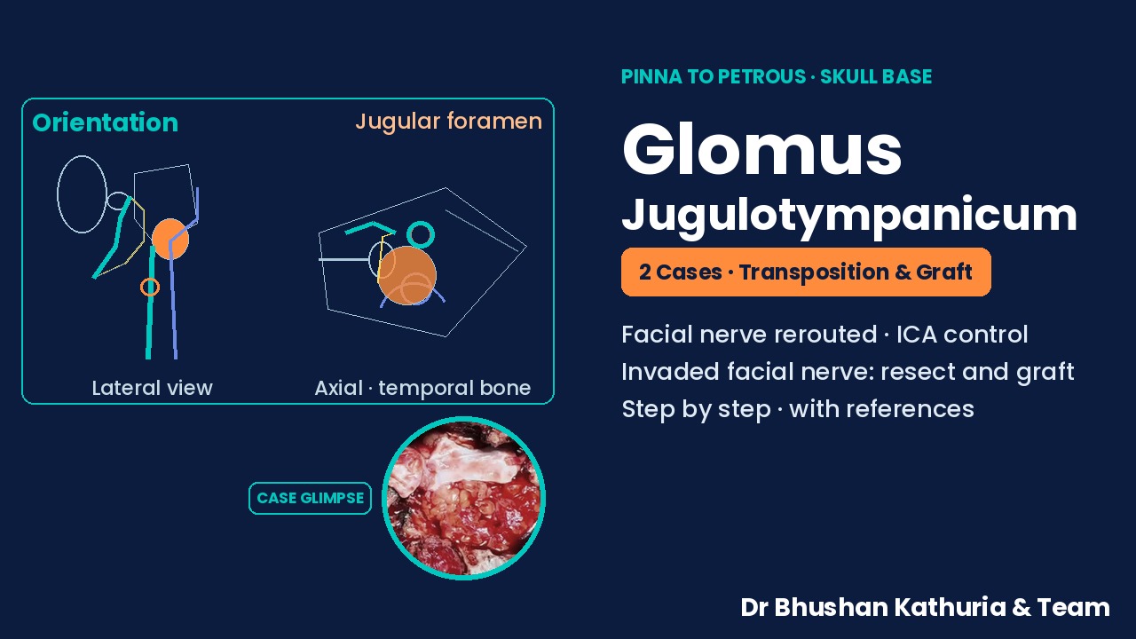
Glomus Jugulotympanicum, Two Cases from Pinna to Petrous: Facial Nerve Transposition with Carotid Control, and Facial Nerve Resection with Interposition Grafting
Two glomus jugulotympanicum cases: facial nerve transposition and facial nerve grafting
Case reportAll surgical case reports → · Skull Base · Sinusitis · Videos · YouTube channel · WhatsApp channel