
Segmental Tracheal Resection and End-to-End Anastomosis for Grade III Post-tracheostomy Tracheal Stenosis
Tracheal resection and primary anastomosis after failed decannulation. Intended audience: ENT residents, airway, head and neck and thoracic surgeons.
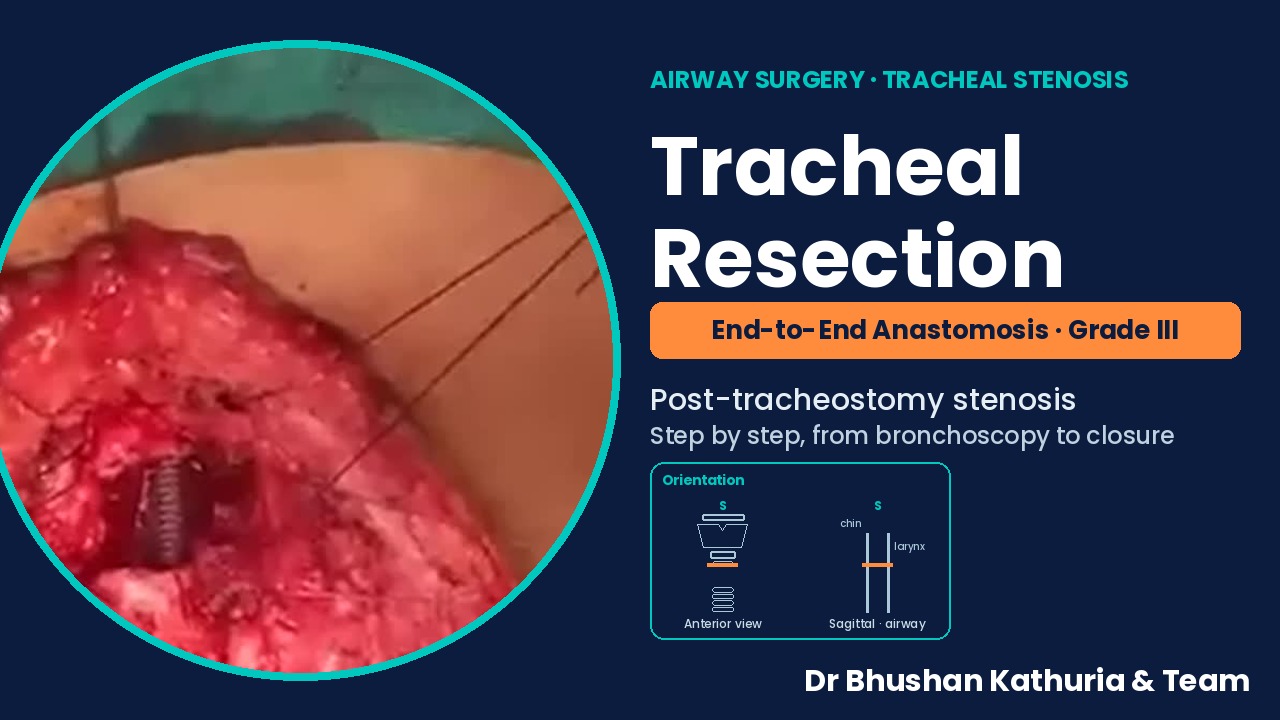
- Diagnosis
- Post-tracheostomy tracheal stenosis, Myer–Cotton grade III
- Procedure
- Segmental tracheal resection with end-to-end anastomosis
- Airway history
- Tracheostomy for 2.5 years; four failed decannulation attempts
- Outcome shown
- Extubated at the end of surgery; decannulated airway
- Video
- 3 min · 12 operative steps
Operative video with step captions and geometric orientation markings. Summary video.
Abstract
A 19-year-old who had been tracheostomised for 2.5 years during management of Japanese encephalitis presented after four unsuccessful attempts at decannulation. Laryngobronchoscopy showed extensive stomal granulation and more than 70% luminal occlusion about 2.5 cm below the vocal cords (Myer–Cotton grade III); CT showed the stenotic segment beginning above the stoma and extending over two-thirds of the tracheal lumen. Through a horizontal neck-crease incision including the stoma, the stenotic segment was resected en bloc with the stoma and granulation tissue, and airway continuity was restored by primary end-to-end anastomosis with interrupted 2-0 polyglactin sutures. A mentosternal suture was placed and the patient was extubated at the end of surgery. The operative sequence is presented step by step with geometric orientation.
Keywords: tracheal stenosis; post-tracheostomy stenosis; tracheal resection; end-to-end anastomosis; failed decannulation; Myer-Cotton grading; airway reconstruction
Introduction
Tracheal stenosis is a recognised complication of prolonged intubation and tracheostomy, arising from mucosal ischaemia, stomal granulation and cartilage collapse at the stoma or cuff site. Endoscopic grading by the Myer–Cotton system (percentage of luminal obstruction) guides management [1]. Mature, circumferential stenosis of grade III or more seldom responds durably to dilatation, and segmental resection with primary end-to-end anastomosis remains the definitive treatment, with high success rates in experienced series [2].
Case presentation
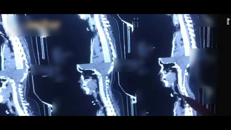
A 19-year-old had undergone tracheostomy 2.5 years earlier during management of Japanese encephalitis. Four attempts at decannulation elsewhere had failed. Laryngobronchoscopy showed extensive granulation at the stoma and more than 70% occlusion of the lumen about 2.5 cm below the vocal cords (Myer–Cotton grade III). CT of the neck showed the stenotic segment beginning above the stoma and extending over two-thirds of the tracheal lumen. Segmental tracheal resection with end-to-end anastomosis was planned. Written informed consent for surgery and for de-identified educational use of images and video was obtained.
Surgical technique
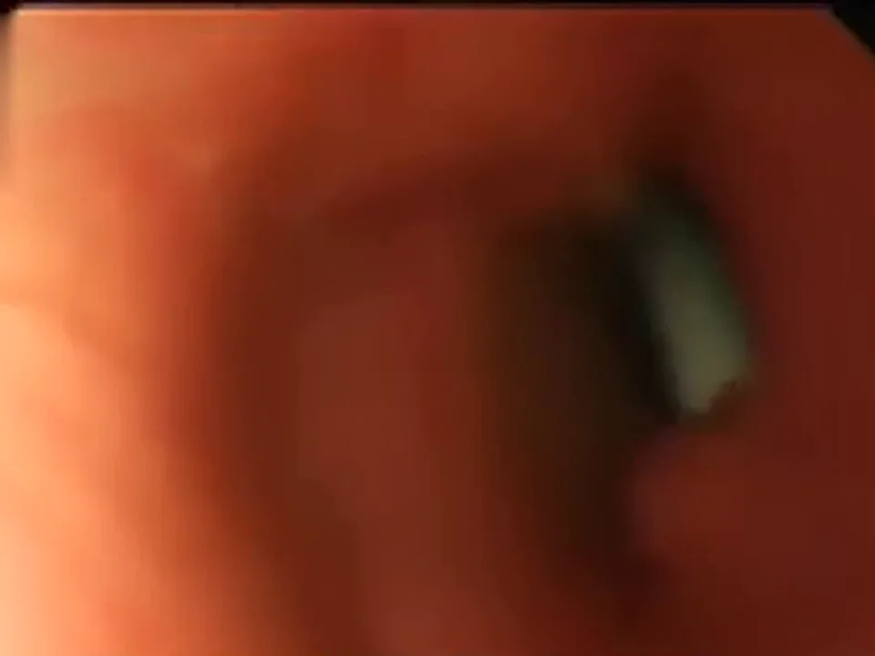
Pre-operative laryngobronchoscopy
Grade III stenosis with more than 70% occlusion about 2.5 cm below the vocal cords; the length and level are confirmed.
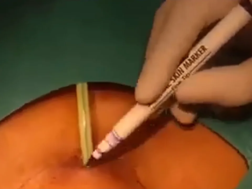
Pre-operative laryngobronchoscopy Skin marking
A horizontal neck-crease incision is marked to include the stoma and the surrounding granulation.
Skin marking Incision and subplatysmal flaps
Superior and inferior subplatysmal flaps are elevated.

Incision and subplatysmal flaps Stomal granulation excised
Granulation tissue at the stoma is dissected out.
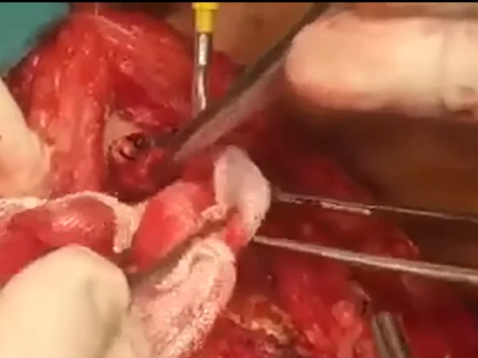
Stomal granulation excised Exposure of the airway
Strap muscles are separated in the midline; the trachea and laryngeal framework are skeletonised.
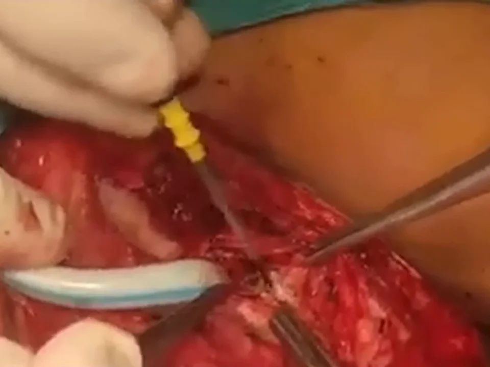
Exposure of the airway Resection of the stenotic segment
The trachea is divided through normal rings above and below the stenosis; the stoma and granulation are excised en bloc.
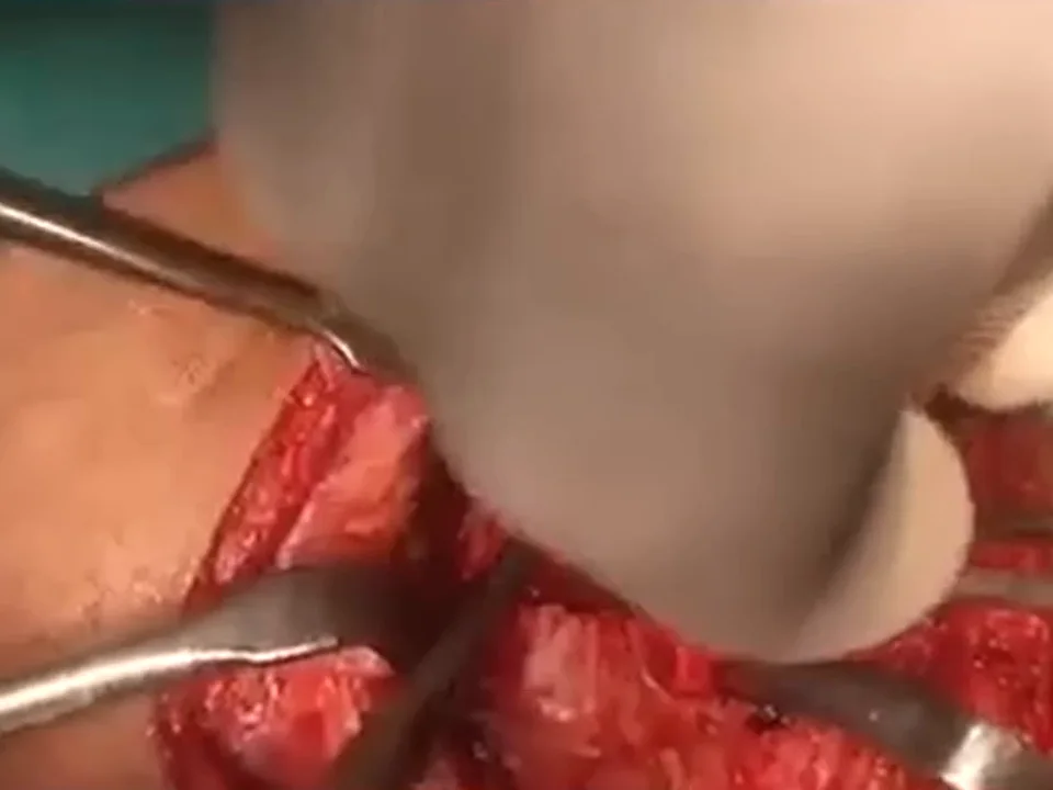
Resection of the stenotic segment Airway control
Ventilation through the stoma is replaced by an orotracheal tube passed across the gap.
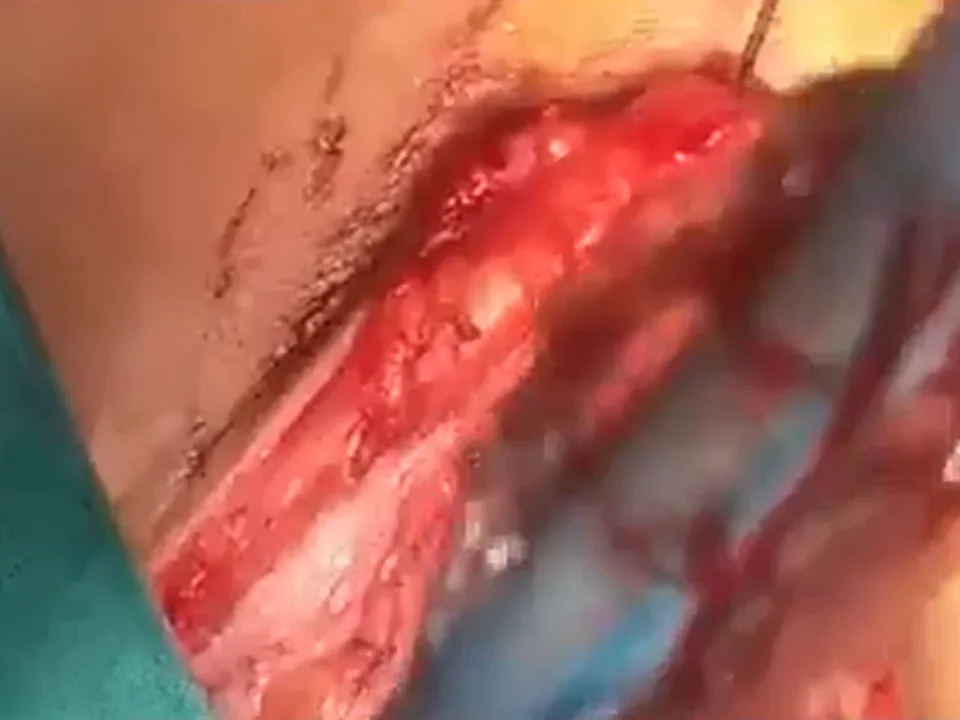
Airway control Tracheal mobilisation
The distal trachea is mobilised and the ends approximated without tension.
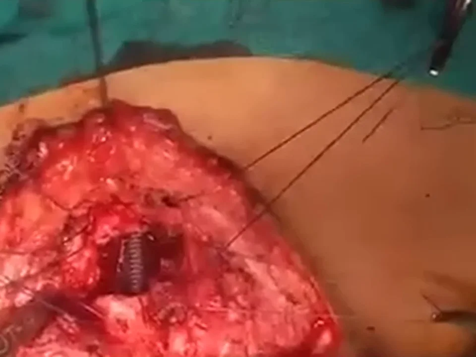
Tracheal mobilisation Anastomosis sutures
Interrupted 2-0 polyglactin (Vicryl) sutures are placed posterolaterally and anteriorly, all before tying.
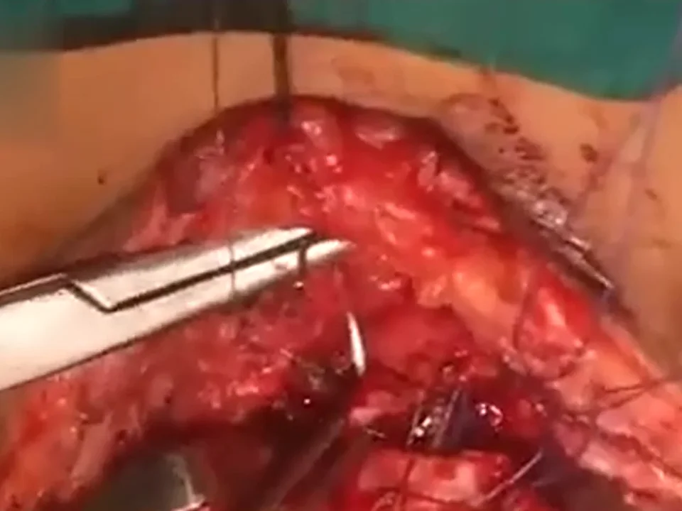
Anastomosis sutures Sutures tied
The tracheal ends are approximated and the anastomosis is confirmed airtight.
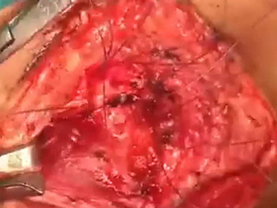
Sutures tied Closure in layers
A drain is placed; strap muscles, deep fascia and platysma are approximated.
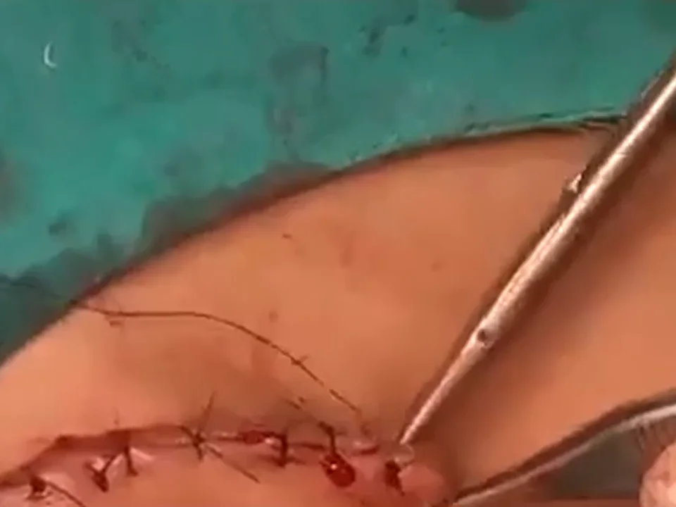
Closure in layers Mentosternal suture
A chin-to-chest suture prevents neck extension and tension on the anastomosis; the patient is extubated at the end of surgery.
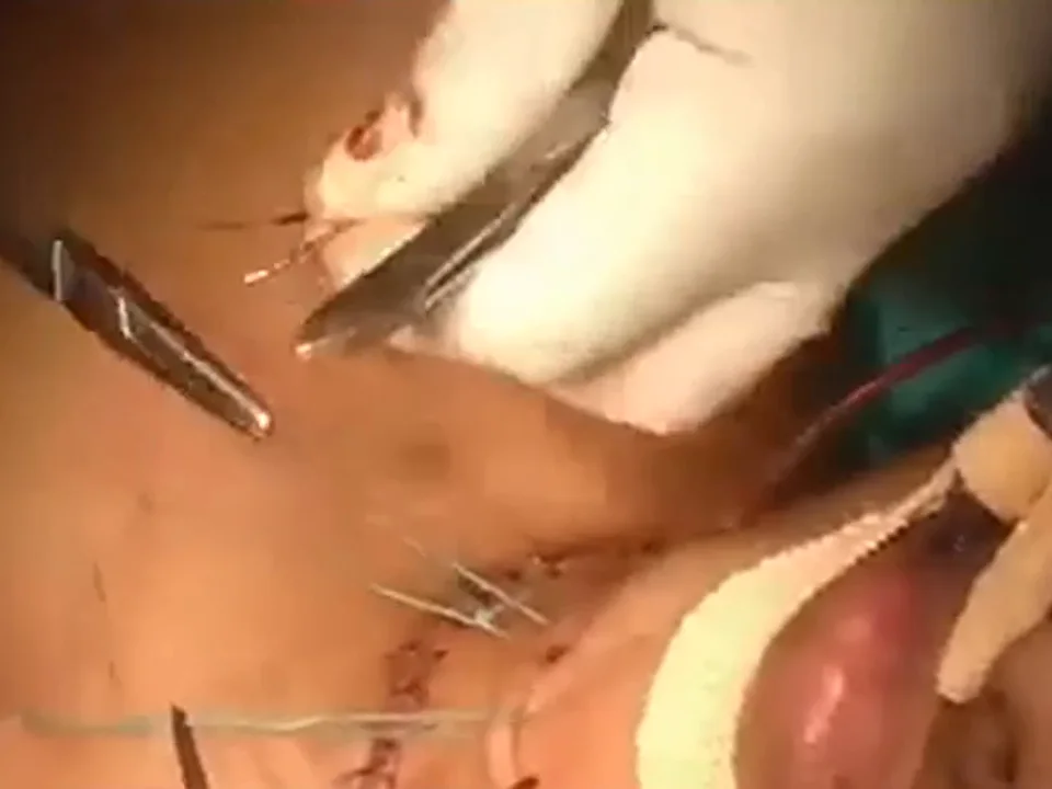
Mentosternal suture
Postoperative management
- Mentosternal suture maintained to keep the neck flexed.
- Humidification, chest physiotherapy and early mobilisation.
- Endoscopic review of the anastomosis before discharge and on follow-up.
- Decannulated airway; surveillance for granulation or re-stenosis.
Discussion
Successful tracheal resection depends on accurate pre-operative definition of the stenosis — its grade, length and distance from the glottis — by endoscopy and cross-sectional imaging. Resection must extend to healthy rings and include the stoma and granulation tissue, since residual diseased cartilage predisposes to re-stenosis. Anastomotic tension is the principal cause of dehiscence and restenosis; it is minimised by limited circumferential mobilisation that preserves the lateral blood supply, by placing all sutures before tying, and by maintaining neck flexion in the early post-operative period [2,3]. Extubation at the end of the procedure avoids a tube across a fresh anastomosis.
Learning points
- Grade the stenosis endoscopically and define its length on CT before planning.
- Resect to healthy rings; include the stoma and granulation tissue.
- Place all anastomotic sutures before tying and avoid tension: mobilisation and neck flexion.
References
- Myer CM 3rd, O'Connor DM, Cotton RT. Proposed grading system for subglottic stenosis based on endotracheal tube sizes. Ann Otol Rhinol Laryngol. 1994;103(4 Pt 1):319-323.
- Grillo HC, Donahue DM, Mathisen DJ, Wain JC, Wright CD. Postintubation tracheal stenosis. Treatment and results. J Thorac Cardiovasc Surg. 1995;109(3):486-492.
- Wright CD, Grillo HC, Wain JC, et al. Anastomotic complications after tracheal resection: prognostic factors and management. J Thorac Cardiovasc Surg. 2004;128(5):731-739.
Consent and ethics
Written informed consent was obtained for surgery and for de-identified use of clinical images and video for education. No identifying information is shown; eyes are blurred where visible. This report is for medical education and does not replace individual clinical advice.
Surgical team
Dr Bhushan Kathuria & Team, Santosh Advance ENT Healthcare, Hisar. Consultants: Dr Bhushan Kathuria, Dr Parmod Jangra, Dr Harshvardhan. Clinical fellows: Dr Ayush, Dr Iqrorjon. Team · Fellowship and previous fellows.
How to cite
Kathuria B, and Team. Segmental Tracheal Resection and End-to-End Anastomosis for Grade III Post-tracheostomy Tracheal Stenosis. Surgical Perspective · Surgical Case Reports. 2026. Available at: https://bhushankathuria.com/case-post-tracheostomy-tracheal-stenosis-resection-anastomosis.html
More case reports
Extended Total Maxillectomy with Infratemporal Fossa, Transpterygoid and Anterior Skull Base Clearance for Sinonasal Mucormycosis
Upfront radical resection for fulminant sinonasal mucormycosis
▶ Video + case report
High-Grade Mucoepidermoid Carcinoma of the Parotid with Facial Nerve and Skin Involvement: Radical Parotidectomy, Radical Neck Dissection and PMMC Flap Reconstruction
Composite parotid resection with single-stage PMMC reconstruction
▶ Video + case report
Giant Pilomatricoma of the Neck Misdiagnosed as Tubercular Lymphadenitis: Wide Local Excision and Two-Stage Deltopectoral Flap Reconstruction
Pilomatricoma presenting as a non-healing neck mass
▶ Video + case report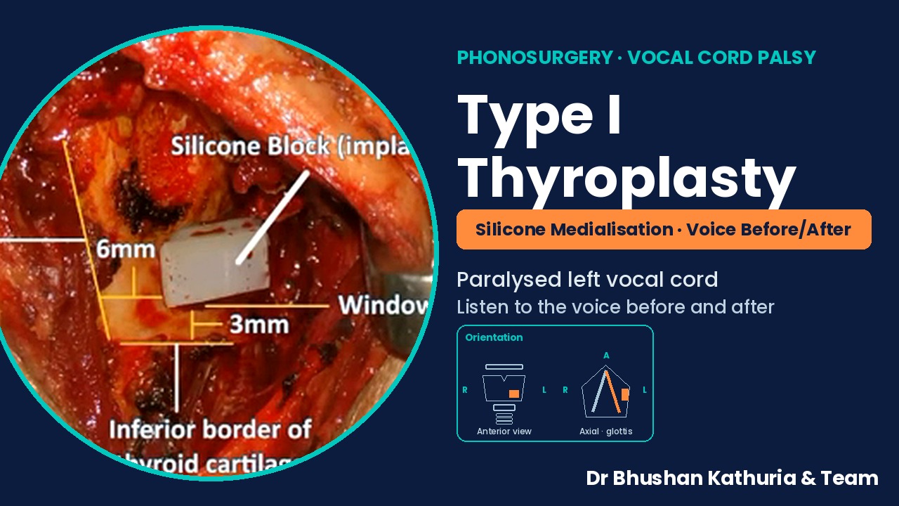
Type I Thyroplasty: Silicone Medialisation of a Paralysed Left Vocal Cord, with Pre- and Post-operative Voice
Medialisation thyroplasty for unilateral vocal fold palsy
▶ Video + case report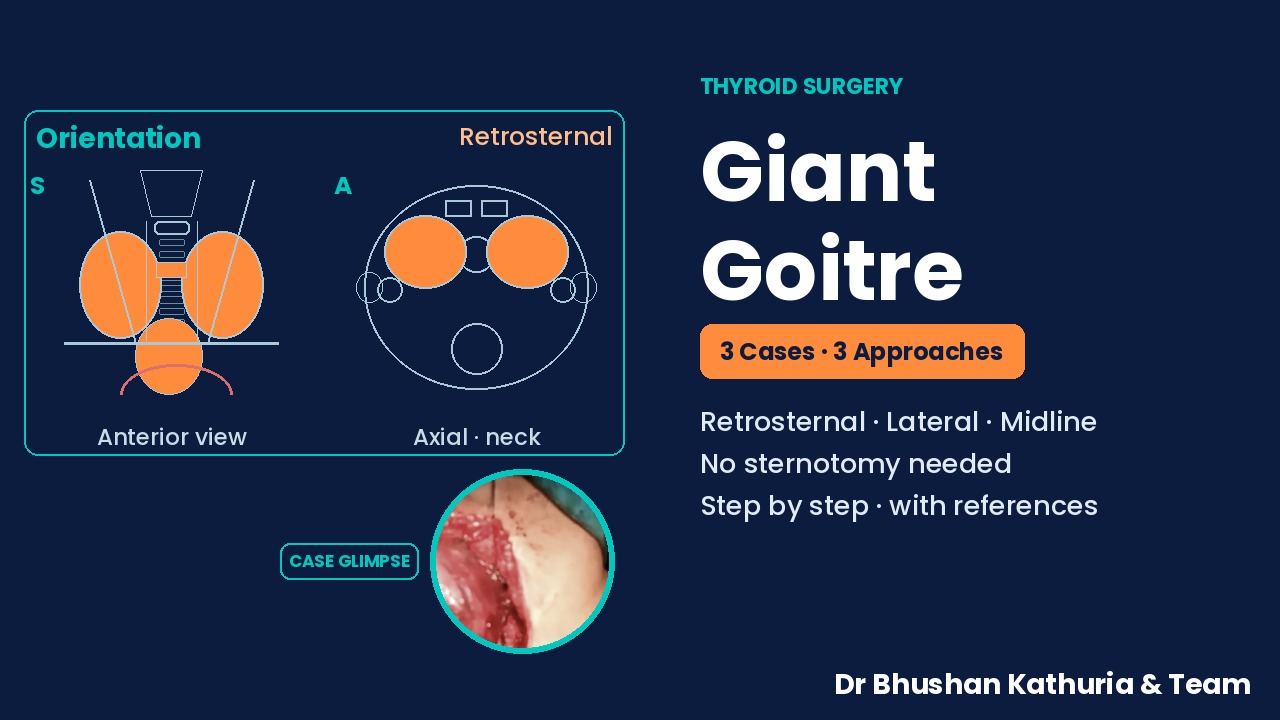
Giant Goitre: Three Cases and Three Approaches — Cervical Delivery of Retrosternal Extension, Lateral Strap-Sparing and Midline Strap-Dividing Thyroidectomy
Three giant goitres, three surgical approaches
Case report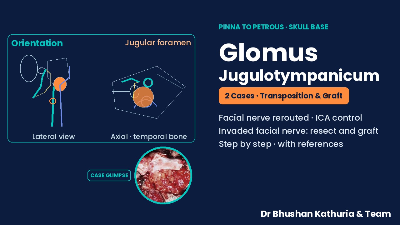
Glomus Jugulotympanicum, Two Cases from Pinna to Petrous: Facial Nerve Transposition with Carotid Control, and Facial Nerve Resection with Interposition Grafting
Two glomus jugulotympanicum cases: facial nerve transposition and facial nerve grafting
Case reportAll surgical case reports → · Voice Swallowing Breathing · Head Neck Cancer · Videos · YouTube channel · WhatsApp channel