
Giant Pilomatricoma of the Neck Misdiagnosed as Tubercular Lymphadenitis: Wide Local Excision and Two-Stage Deltopectoral Flap Reconstruction
Pilomatricoma presenting as a non-healing neck mass. Intended audience: ENT, head and neck, plastic and general surgeons.
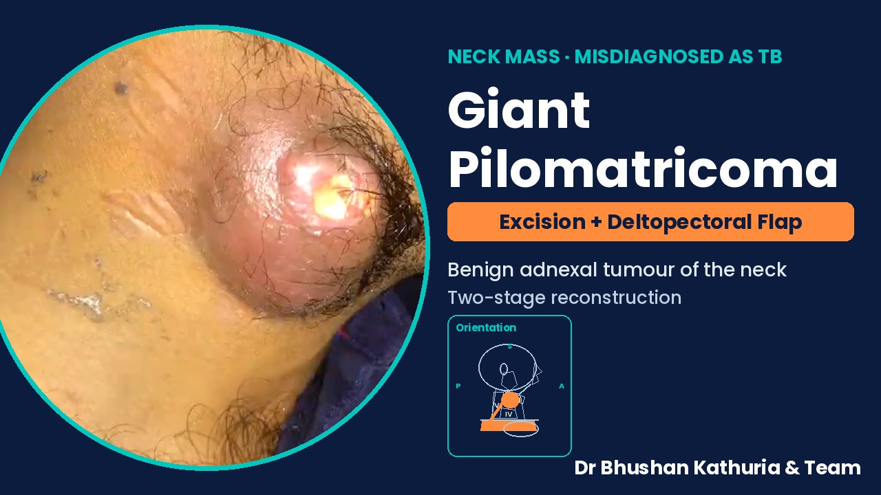
- Diagnosis
- Pilomatricoma of the neck (misdiagnosed as TB)
- Procedure
- Wide local excision with skin
- Defect
- About 8 × 7.5 cm
- Reconstruction
- Deltopectoral flap, divided at day 21
- Video
- 2 min · step-by-step
Operative video with step captions and geometric orientation markings. Summary video.
Abstract
Pilomatricoma is a benign adnexal tumour of hair-matrix origin, most often in the head and neck, and frequently misdiagnosed clinically. We describe a young adult treated empirically for tubercular lymphadenitis for six months, in whom a 7 × 6.5 cm right neck mass with a discharging sinus proved on biopsy to be pilomatricoma. Wide local excision with the involved skin left an 8 × 7.5 cm defect, reconstructed with a medially based deltopectoral flap and divided at three weeks.
Keywords: pilomatricoma; pilomatrixoma; neck mass; tubercular lymphadenitis; deltopectoral flap
Introduction
Pilomatricoma (calcifying epithelioma of Malherbe) usually presents as a firm, slow-growing subcutaneous nodule and is easily confused with other skin and neck masses [1]. In regions where tuberculosis is endemic, a non-healing neck mass may be treated empirically without tissue diagnosis.
Case presentation
A 21-year-old man had a right neck swelling for six months, treated as tubercular granuloma without response. The mass enlarged progressively with skin breakdown and a discharging sinus. Biopsy showed pilomatricoma.
Written informed consent for surgery and for de-identified educational use of images was obtained.
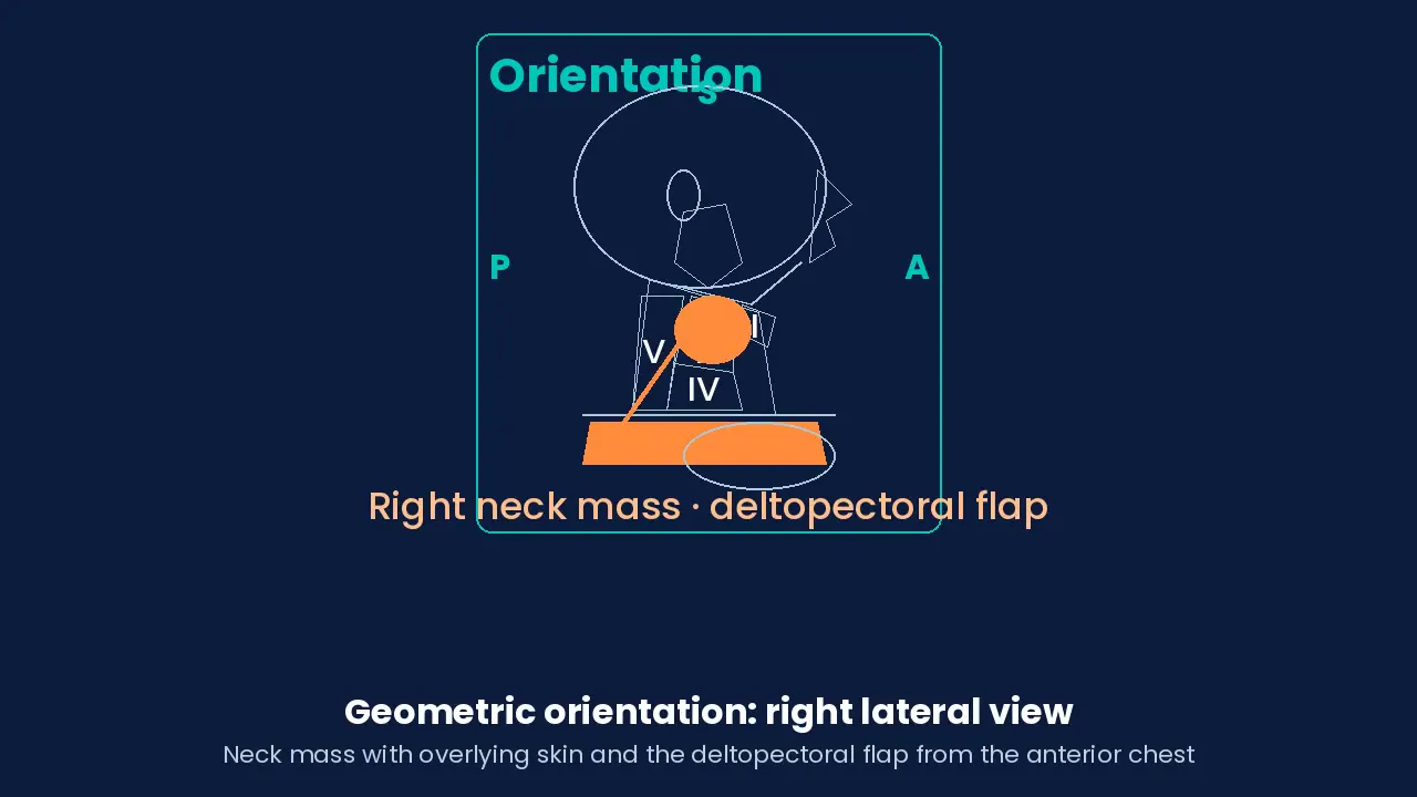
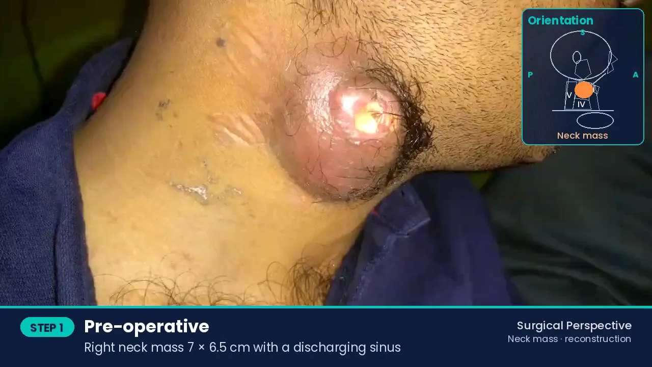
Surgical technique
Marking with a safe margin
Elliptical skin excision around the sinus and involved skin.
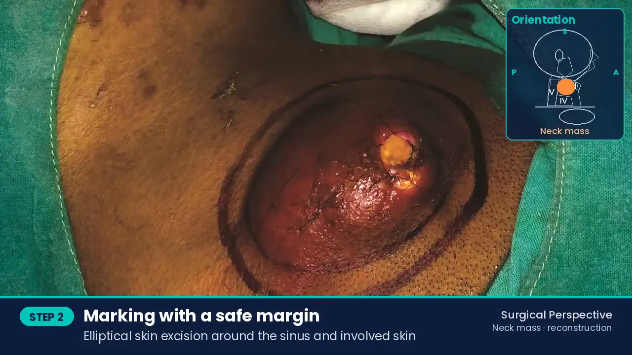
Marking with a safe margin Wide local excision
Mass removed en bloc with the overlying skin.
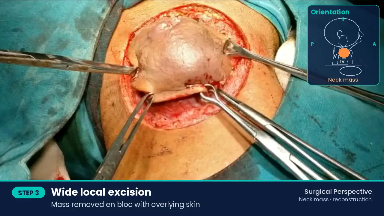
Wide local excision Defect and specimen
Defect about 8 × 7.5 cm, too large for primary closure.
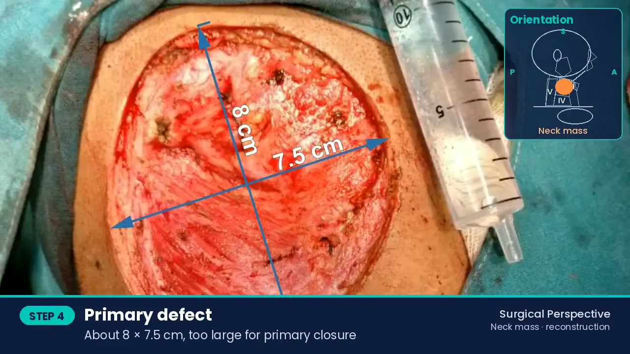
Defect and specimen Deltopectoral flap
Medially based on internal mammary perforators, raised in the subfascial plane from lateral to medial and transposed to the neck; donor site closed primarily [2].
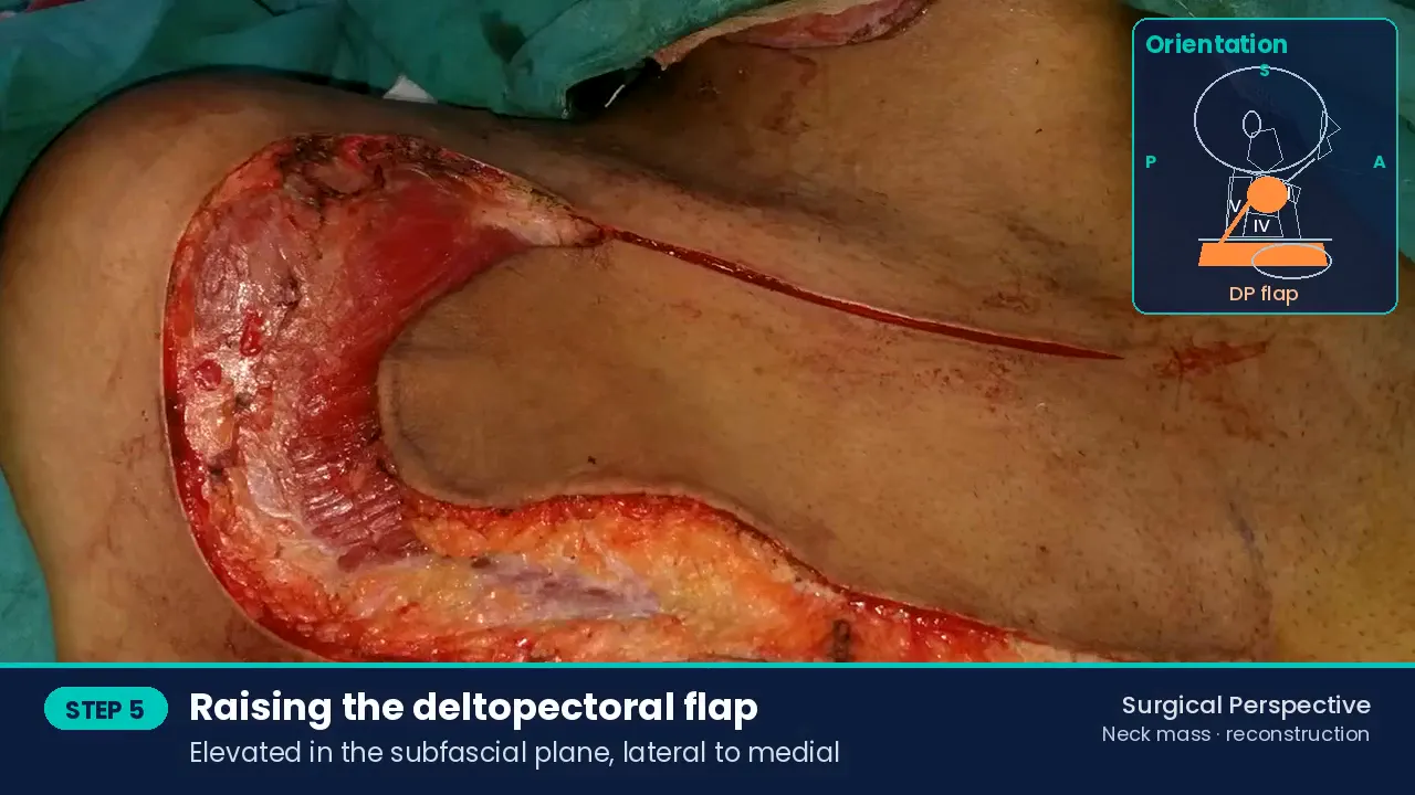
Deltopectoral flap Flap inset
Defect covered.
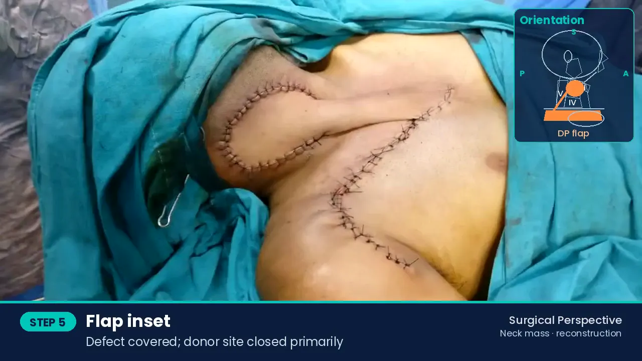
Flap inset Second stage
Flap division at day 21 after neovascularisation from the recipient bed; the unused pedicle returned to the chest.
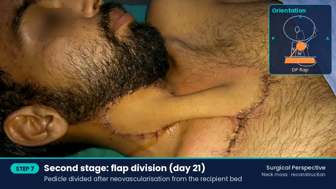
Second stage Final result
Stable cover with acceptable contour.
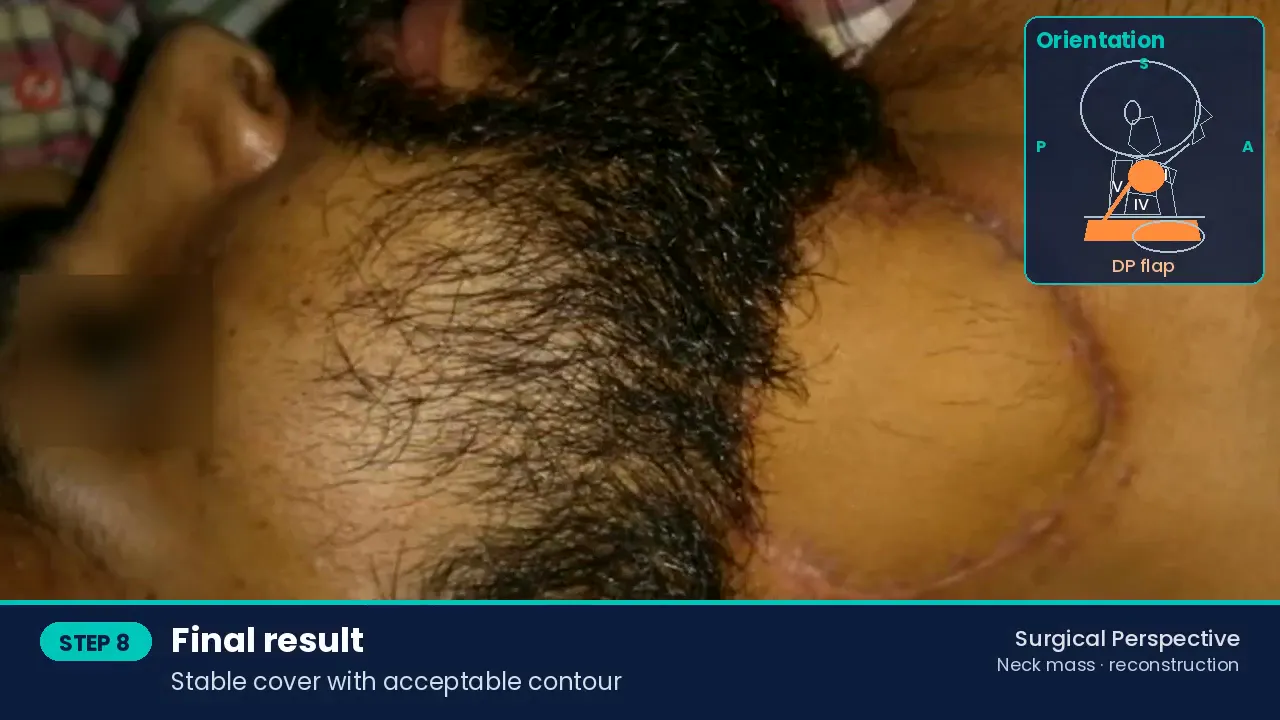
Final result
Postoperative management
- Wound care and flap monitoring; second-stage division at three weeks.
- Histopathology of the excised specimen confirmed the diagnosis.
Discussion
A neck mass that does not respond to antitubercular treatment needs a tissue diagnosis before therapy is prolonged. Complete excision is curative for pilomatricoma, with a low recurrence rate. When excision leaves a large neck skin defect, the deltopectoral flap provides thin, reliable cover.
Learning points
- Not every neck lump is TB: obtain a tissue diagnosis when there is no response to treatment.
- Excise completely with the involved skin and sinus.
- The deltopectoral flap is a reliable two-stage option for large neck skin defects.
References
- Hu JL, Yoo H, Kwon ST, et al. Clinical analysis and review of literature on pilomatrixoma in pediatric patients. Arch Craniofac Surg. 2020;21(5):288-293. https://doi.org/10.7181/acfs.2020.00528
- Bakamjian VY. A two-stage method for pharyngoesophageal reconstruction with a primary pectoral skin flap. Plast Reconstr Surg. 1965;36:173-184.
Consent and ethics
Written informed consent was obtained for surgery and for de-identified use of clinical images and video for education. No identifying information is shown; eyes are blurred where visible. This report is for medical education and does not replace individual clinical advice.
Surgical team
Dr Bhushan Kathuria & Team, Santosh Advance ENT Healthcare, Hisar. Consultants: Dr Bhushan Kathuria, Dr Parmod Jangra, Dr Harshvardhan. Clinical fellows: Dr Ayush, Dr Iqrorjon. Team · Fellowship and previous fellows.
How to cite
Kathuria B, and Team. Giant Pilomatricoma of the Neck Misdiagnosed as Tubercular Lymphadenitis: Wide Local Excision and Two-Stage Deltopectoral Flap Reconstruction. Surgical Perspective · Surgical Case Reports. 2026. Available at: https://bhushankathuria.com/case-pilomatricoma-neck-deltopectoral-flap.html
More case reports

Extended Total Maxillectomy with Infratemporal Fossa, Transpterygoid and Anterior Skull Base Clearance for Sinonasal Mucormycosis
Upfront radical resection for fulminant sinonasal mucormycosis
▶ Video + case report
High-Grade Mucoepidermoid Carcinoma of the Parotid with Facial Nerve and Skin Involvement: Radical Parotidectomy, Radical Neck Dissection and PMMC Flap Reconstruction
Composite parotid resection with single-stage PMMC reconstruction
▶ Video + case report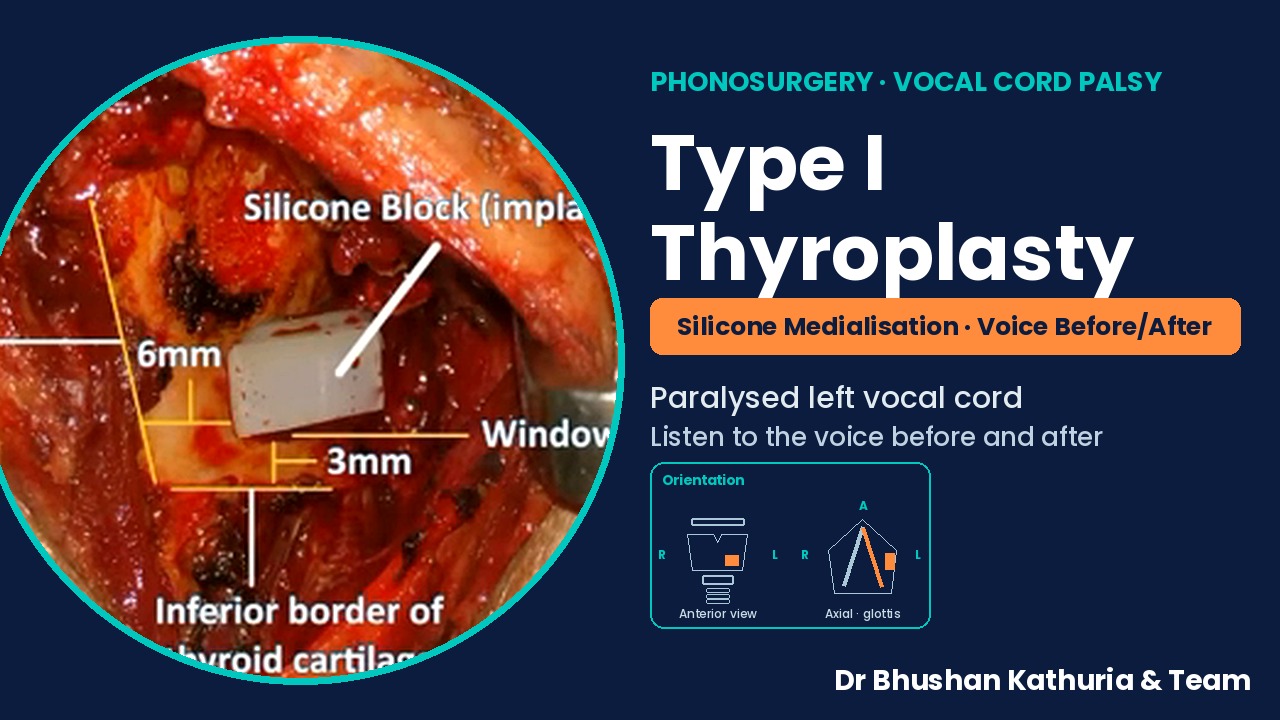
Type I Thyroplasty: Silicone Medialisation of a Paralysed Left Vocal Cord, with Pre- and Post-operative Voice
Medialisation thyroplasty for unilateral vocal fold palsy
▶ Video + case report
Segmental Tracheal Resection and End-to-End Anastomosis for Grade III Post-tracheostomy Tracheal Stenosis
Tracheal resection and primary anastomosis after failed decannulation
▶ Video + case report
Giant Goitre: Three Cases and Three Approaches — Cervical Delivery of Retrosternal Extension, Lateral Strap-Sparing and Midline Strap-Dividing Thyroidectomy
Three giant goitres, three surgical approaches
Case report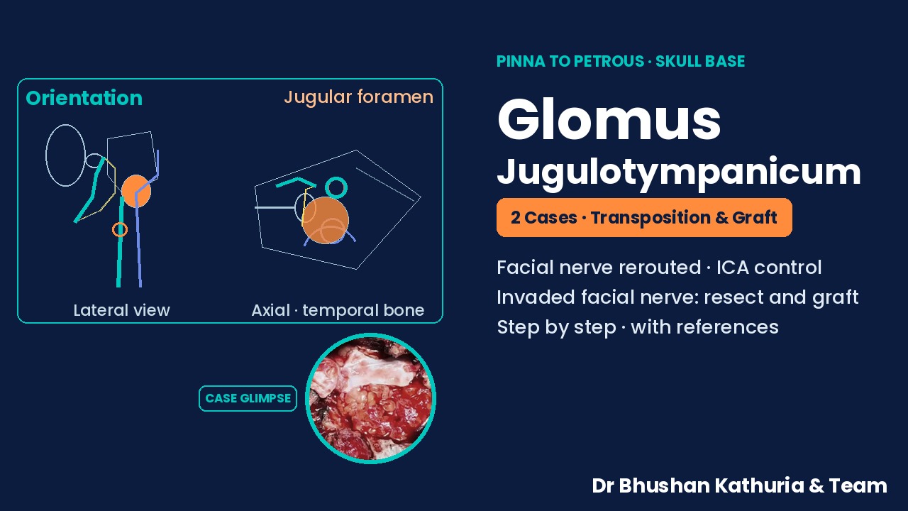
Glomus Jugulotympanicum, Two Cases from Pinna to Petrous: Facial Nerve Transposition with Carotid Control, and Facial Nerve Resection with Interposition Grafting
Two glomus jugulotympanicum cases: facial nerve transposition and facial nerve grafting
Case reportAll surgical case reports → · Head Neck Cancer · Videos · YouTube channel · WhatsApp channel