Mastering Precision in Otorhinolaryngology
Companion manual · DissectionWithin the
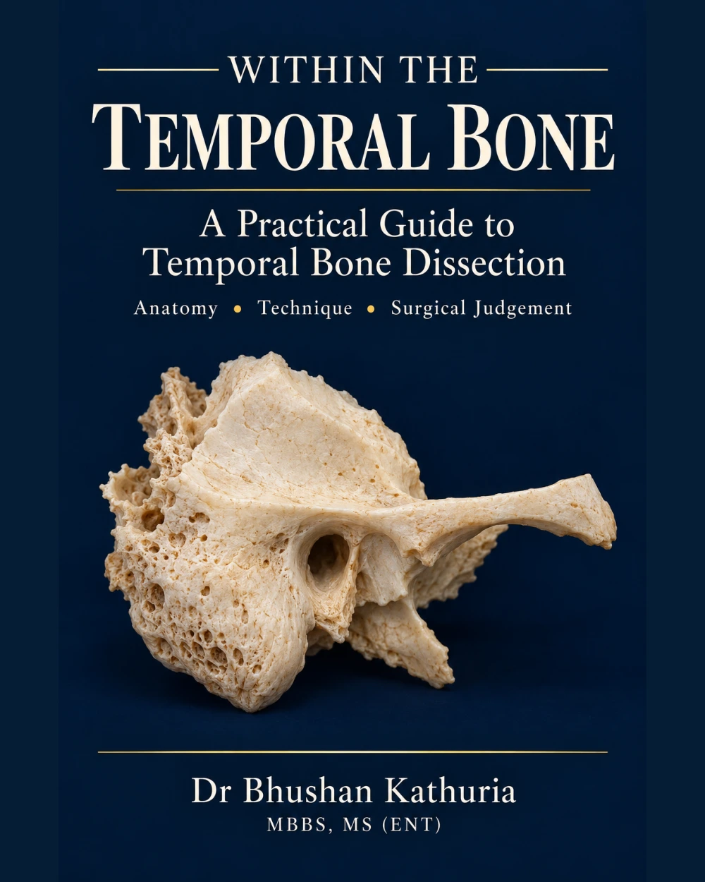
Temporal Bone
A Practical Guide to Temporal Bone Dissection
A surgeon’s companion to temporal bone dissection, from laboratory preparation to selected lateral skull-base exposures.
232 pages in the PDF · 17 chapters · atlas illustrations · 67 real-image pages · 20 companion videos
Free to read online · no registration or payment.
Read, orient, reflect
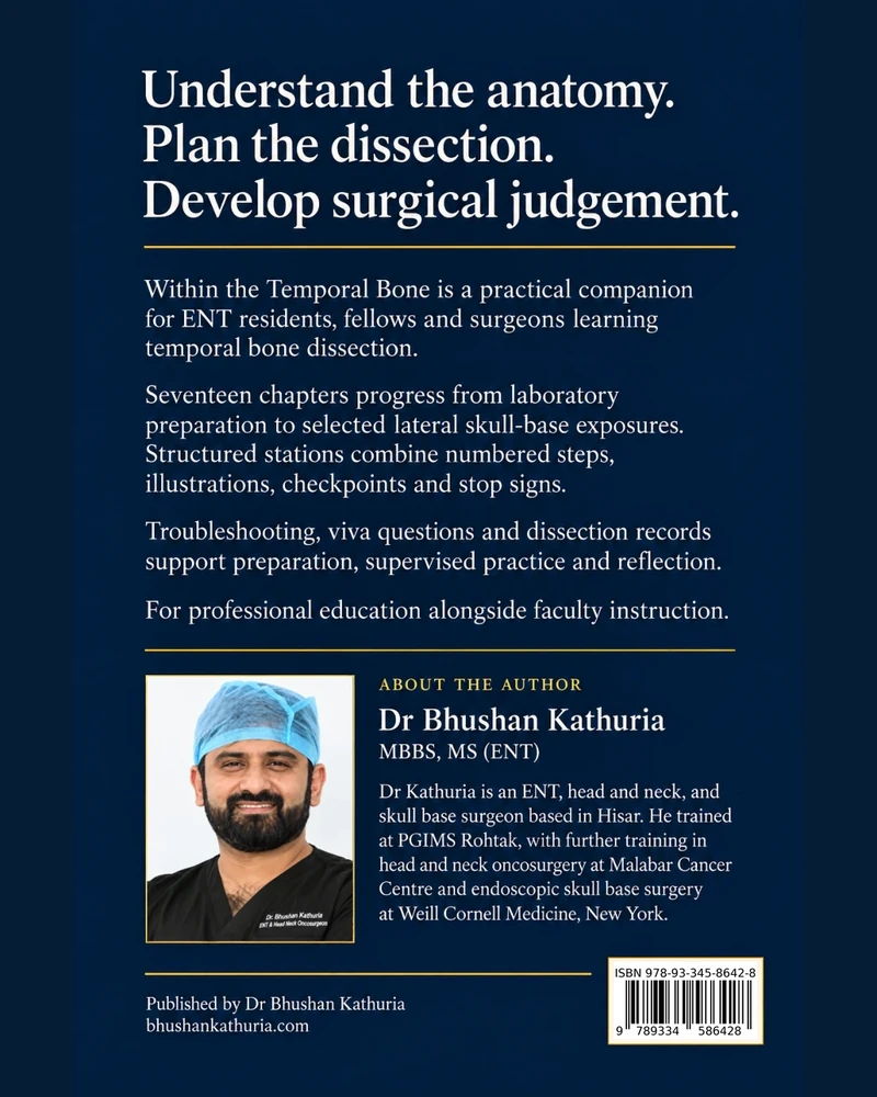
Written for residents, fellows and surgeons, this manual breaks every dissection into stations, each with numbered steps, a figure, a checkpoint and a stop sign. Each chapter adds fault finding, viva questions with answers and a debrief; the appendices hold a dissection record, terminology, photocopiable score sheets and an index. It is intended for professional education alongside instruction and local protocols.
Publication details revised 29 October 2026. Includes the author’s updated acknowledgements, corrected publisher address, and AI assistance disclosure. Dr S. P. S. Yadav’s specialist-reviewer credit is retained from the source edition. Revised 15 November 2026: the drawings in the station panels and the captioned figures are replaced with credited real images (real dissections, classic anatomical plates, real CT and operative photographs; source and licence printed with each), and the chapters close with “Real images” pages. The AI-generated orientation image was withdrawn.
AI assistance disclosure
Generative artificial intelligence tools assisted with drafting, editing and organising portions of the text, and with generating or modifying illustrations. AI-assisted material may contain factual, anatomical or visual inaccuracies. Illustrations are schematic educational aids, are not to scale, and should not be treated as operative references. Readers should verify information against authoritative sources, specimen anatomy and supervised specialist instruction. Responsibility for the published content remains with the author.
Read online
Browse contents and chapters
- Cover
- Publication details & AI disclosure
- Acknowledgements
- Chapter 1
- Plate · Chapter 1 · Real images · 1 of 2
- Plate · Chapter 1 · Real images · 2 of 2
- Chapter 2
- Plate · Chapter 2 · Real images · 1 of 2
- Plate · Chapter 2 · Real images · 2 of 2
- Chapter 3
- Plate · Chapter 3 · From the open atlas
- Plate · Chapter 3 · Real images · 1 of 4
- Plate · Chapter 3 · Real images · 2 of 4
- Plate · Chapter 3 · Real images · 3 of 4
- Plate · Chapter 3 · Real images · 4 of 4
- Chapter 4
- Plate · Chapter 4 · Real images · 1 of 4
- Plate · Chapter 4 · Real images · 2 of 4
- Plate · Chapter 4 · Real images · 3 of 4
- Plate · Chapter 4 · Real images · 4 of 4
- Chapter 5
- Plate · Chapter 5 · Real images · 1 of 8
- Plate · Chapter 5 · Real images · 2 of 8
- Plate · Chapter 5 · Real images · 3 of 8
- Plate · Chapter 5 · Real images · 4 of 8
- Plate · Chapter 5 · Real images · 5 of 8
- Plate · Chapter 5 · Real images · 6 of 8
- Plate · Chapter 5 · Real images · 7 of 8
- Plate · Chapter 5 · Real images · 8 of 8
- Chapter 6
- Plate · Chapter 6 · In the operating theatre
- Plate · Chapter 6 · From the open atlas
- Chapter 7
- Plate · Chapter 7 · From the open atlas
- Plate · Chapter 7 · Real images · 1 of 3
- Plate · Chapter 7 · Real images · 2 of 3
- Plate · Chapter 7 · Real images · 3 of 3
- Chapter 8
- Plate · Chapter 8 · From the dissection hall
- Plate · Chapter 8 · From the open atlas
- Plate · Chapter 8 · Real images · 1 of 2
- Plate · Chapter 8 · Real images · 2 of 2
- Chapter 9
- Plate · Chapter 9 · In the operating theatre
- Plate · Chapter 9 · Real images
- Chapter 10
- Plate · Chapter 10 · From the dissection hall
- Plate · Chapter 10 · From the open atlas
- Plate · Chapter 10 · Real images · 1 of 2
- Plate · Chapter 10 · Real images · 2 of 2
- Chapter 11
- Plate · Chapter 11 · Real images · 1 of 4
- Plate · Chapter 11 · Real images · 2 of 4
- Plate · Chapter 11 · Real images · 3 of 4
- Plate · Chapter 11 · Real images · 4 of 4
- Chapter 12
- Plate · Chapter 12 · Real images · 1 of 4
- Plate · Chapter 12 · Real images · 2 of 4
- Plate · Chapter 12 · Real images · 3 of 4
- Plate · Chapter 12 · Real images · 4 of 4
- Chapter 13
- Plate · Chapter 13 · Orientation
- Plate · Chapter 13 · From the open atlas
- Plate · Chapter 13 · Real images · 1 of 3
- Plate · Chapter 13 · Real images · 2 of 3
- Plate · Chapter 13 · Real images · 3 of 3
- Plate · Chapter 13 · The facial nerve, complete
- Plate · Chapter 13 · The facial nerve, complete
- Plate · Chapter 13 · The facial nerve, complete
- Plate · Chapter 13 · The facial nerve, complete
- Plate · Chapter 13 · The facial nerve, complete
- Chapter 14
- Plate · Chapter 14 · From the open atlas
- Plate · Chapter 14 · Real images · 1 of 5
- Plate · Chapter 14 · Real images · 2 of 5
- Plate · Chapter 14 · Real images · 3 of 5
- Plate · Chapter 14 · Real images · 4 of 5
- Plate · Chapter 14 · Real images · 5 of 5
- Chapter 15
- Plate · Chapter 15 · Real images · 1 of 3
- Plate · Chapter 15 · Real images · 2 of 3
- Plate · Chapter 15 · Real images · 3 of 3
- Chapter 16
- Plate · Chapter 16 · Real images · 1 of 2
- Plate · Chapter 16 · Real images · 2 of 2
- Chapter 17
- Plate · Chapter 17 · Real images
From the dissection hall and the operating theatre
10 pages use real frames from the GeNext MedEd Foundation cadaveric dissection series (Dr Raman Sharma, Dr Vinit Panchal) and from live cochlear implant surgery by Dr Bhushan Kathuria & Team, with a QR code to the full video; patient identity is masked. 7 further pages carry labelled photographs from The Open Access Atlas of Otolaryngology, Head & Neck Operative Surgery (ed. J Fagan, University of Cape Town; chapters by T Harris, T Linder, R Mathew and P Patel), used under CC BY-NC 3.0. 50 “Real images” pages close the chapters with real dissections from the Bassett Collection (D. L. Bassett, A Stereoscopic Atlas of Human Anatomy, Lane Medical Library, Stanford University, CC BY-SA 4.0), classic plates from Gray’s Anatomy (1918) and Burghard’s A System of Operative Surgery (1909) (public domain), labelled CT from Radiopaedia.org (case courtesy of Craig Hacking, CC BY-NC-SA 3.0), operative photographs from the Open Access Atlas, and specimen and equipment photographs from Wikimedia Commons; each image carries its own source and licence. Each page sits in its chapter in the reader and the PDF.
 Chapter 06 · In the operating theatreCortical Mastoidectomy
 Chapter 08 · From the dissection hallThe Facial Recess and Posterior Tympanotomy
 Chapter 09 · In the operating theatreThe Round Window and Cochlear Implant Corridor
 Chapter 10 · From the dissection hallCanal Wall Down Dissection and Cavity Design
 Chapter 13 · OrientationThe Intratemporal Facial Nerve
 Chapter 03 · From the open atlasOrientation and Surface Anatomy
 Chapter 06 · From the open atlasCortical Mastoidectomy
 Chapter 07 · From the open atlasThe Antrum, Aditus and Epitympanum
 Chapter 08 · From the open atlasThe Facial Recess and Posterior Tympanotomy
 Chapter 10 · From the open atlasCanal Wall Down Dissection and Cavity Design
 Chapter 13 · From the open atlasThe Intratemporal Facial Nerve
 Chapter 14 · From the open atlasThe Labyrinth and Endolymphatic Sac Region
 Chapter 01 · Real images · 1 of 2Laboratory Preparation and Specimen Care
 Chapter 01 · Real images · 2 of 2Laboratory Preparation and Specimen Care
 Chapter 02 · Real images · 1 of 2The Microscope, Drill and Microinstruments
 Chapter 02 · Real images · 2 of 2The Microscope, Drill and Microinstruments
 Chapter 03 · Real images · 1 of 4Orientation and Surface Anatomy
 Chapter 03 · Real images · 2 of 4Orientation and Surface Anatomy
 Chapter 03 · Real images · 3 of 4Orientation and Surface Anatomy
 Chapter 03 · Real images · 4 of 4Orientation and Surface Anatomy
 Chapter 04 · Real images · 1 of 4Reading Temporal Bone Imaging
 Chapter 04 · Real images · 2 of 4Reading Temporal Bone Imaging
 Chapter 04 · Real images · 3 of 4Reading Temporal Bone Imaging
 Chapter 04 · Real images · 4 of 4Reading Temporal Bone Imaging
 Chapter 05 · Real images · 1 of 8Transcanal Exposure and the Middle Ear
 Chapter 05 · Real images · 2 of 8Transcanal Exposure and the Middle Ear
 Chapter 05 · Real images · 3 of 8Transcanal Exposure and the Middle Ear
 Chapter 05 · Real images · 4 of 8Transcanal Exposure and the Middle Ear
 Chapter 05 · Real images · 5 of 8Transcanal Exposure and the Middle Ear
 Chapter 05 · Real images · 6 of 8Transcanal Exposure and the Middle Ear
 Chapter 05 · Real images · 7 of 8Transcanal Exposure and the Middle Ear
 Chapter 05 · Real images · 8 of 8Transcanal Exposure and the Middle Ear
 Chapter 07 · Real images · 1 of 3The Antrum, Aditus and Epitympanum
 Chapter 07 · Real images · 2 of 3The Antrum, Aditus and Epitympanum
 Chapter 07 · Real images · 3 of 3The Antrum, Aditus and Epitympanum
 Chapter 08 · Real images · 1 of 2The Facial Recess and Posterior Tympanotomy
 Chapter 08 · Real images · 2 of 2The Facial Recess and Posterior Tympanotomy
 Chapter 09 · Real imagesThe Round Window and Cochlear Implant Corridor
 Chapter 10 · Real images · 1 of 2Canal Wall Down Dissection and Cavity Design
 Chapter 10 · Real images · 2 of 2Canal Wall Down Dissection and Cavity Design
 Chapter 11 · Real images · 1 of 4The Ossicular Chain and Reconstruction Principles
 Chapter 11 · Real images · 2 of 4The Ossicular Chain and Reconstruction Principles
 Chapter 11 · Real images · 3 of 4The Ossicular Chain and Reconstruction Principles
 Chapter 11 · Real images · 4 of 4The Ossicular Chain and Reconstruction Principles
 Chapter 12 · Real images · 1 of 4The Stapes and Oval Window
 Chapter 12 · Real images · 2 of 4The Stapes and Oval Window
 Chapter 12 · Real images · 3 of 4The Stapes and Oval Window
 Chapter 12 · Real images · 4 of 4The Stapes and Oval Window
 Chapter 13 · Real images · 1 of 3The Intratemporal Facial Nerve
 Chapter 13 · Real images · 2 of 3The Intratemporal Facial Nerve
 Chapter 13 · Real images · 3 of 3The Intratemporal Facial Nerve
 Chapter 14 · Real images · 1 of 5The Labyrinth and Endolymphatic Sac Region
 Chapter 14 · Real images · 2 of 5The Labyrinth and Endolymphatic Sac Region
 Chapter 14 · Real images · 3 of 5The Labyrinth and Endolymphatic Sac Region
 Chapter 14 · Real images · 4 of 5The Labyrinth and Endolymphatic Sac Region
 Chapter 14 · Real images · 5 of 5The Labyrinth and Endolymphatic Sac Region
 Chapter 15 · Real images · 1 of 3The Internal Auditory Canal and Translabyrinthine Exposure
 Chapter 15 · Real images · 2 of 3The Internal Auditory Canal and Translabyrinthine Exposure
 Chapter 15 · Real images · 3 of 3The Internal Auditory Canal and Translabyrinthine Exposure
 Chapter 16 · Real images · 1 of 2The Middle Fossa and Petrous Apex
 Chapter 16 · Real images · 2 of 2The Middle Fossa and Petrous Apex
 Chapter 17 · Real imagesCompletion, Complications and Transfer to the Theatre
 Chapter 13 · The facial nerve, completeThe Intratemporal Facial Nerve
 Chapter 13 · The facial nerve, completeThe Intratemporal Facial Nerve
 Chapter 13 · The facial nerve, completeThe Intratemporal Facial Nerve
 Chapter 13 · The facial nerve, completeThe Intratemporal Facial Nerve
 Chapter 13 · The facial nerve, completeThe Intratemporal Facial Nerve
Watch the dissections and the operations
20 videos are linked to the chapters they illustrate: cadaveric dissections from GeNext workshops beside live operations. The same links are printed as QR codes in the PDF. Videos are for professional education.
 Chapter 03 · Orientation and Surface AnatomyTemporal Bone: talk by Dr Vikas KakkarHAOICON 2024 · conference talk
 Chapter 05 · Transcanal Exposure and the Middle EarEndoscopic Tympanoplasty: Hearing restorationLive surgery · transcanal access to the middle ear
 Chapter 06 · Cortical MastoidectomyCortical Mastoidectomy, Step by StepLive surgery · see the plate after Chapter 6
 Chapter 07 · The Antrum, Aditus and EpitympanumRecurrent Cholesteatoma: Endoscopic approachLive surgery · attic and epitympanum
 Chapter 08 · The Facial Recess and Posterior TympanotomyPosterior Tympanotomy · Temporal Bone Dissection Part 1Cadaveric dissection · Dr Raman Sharma
 Chapter 08 · The Facial Recess and Posterior TympanotomyCochlear Implant Surgery, Part 2: Posterior TympanotomyLive surgery · the same corridor in a patient
 Chapter 09 · The Round Window and Cochlear Implant CorridorCochlear Implant Surgery, Part 3: Opening the CochleaLive surgery · see the plate after Chapter 9
 Chapter 09 · The Round Window and Cochlear Implant CorridorCochlear Implant Surgery, Part 4: Electrode InsertionLive surgery · microscopic view
 Chapter 10 · Canal Wall Down Dissection and Cavity DesignCanal Wall Down Mastoidectomy · Temporal Bone Dissection Part 2Cadaveric dissection · Dr Raman Sharma

 Chapter 11 · The Ossicular Chain and Reconstruction PrinciplesTympano-Ossiculoplasty: PORP and malleus relocationLive surgery · ossicular reconstruction
 Chapter 12 · The Stapes and Oval WindowOtosclerosis: Endoscopic stapedotomyLive surgery · stapes and oval window
 Chapter 13 · The Intratemporal Facial NerveFacial Nerve Decompression · Temporal Bone Dissection Part 3Cadaveric dissection · Dr Raman Sharma
 Chapter 13 · The Intratemporal Facial NerveFacial Nerve Transposition · Temporal Bone Dissection Part 4Cadaveric dissection · Dr Vinit Panchal
 Chapter 13 · The Intratemporal Facial NerveDigastric Ridge & Stylomastoid Foramen · Dissection Part 5Cadaveric dissection · Dr Vinit Panchal
 Chapter 13 · The Intratemporal Facial NerveCochleariform Process & GSPN · Temporal Bone Dissection Part 6Cadaveric dissection · Dr Vinit Panchal
 Chapter 13 · The Intratemporal Facial NerveRerouting the Facial Nerve · Temporal Bone Dissection Part 7Cadaveric dissection
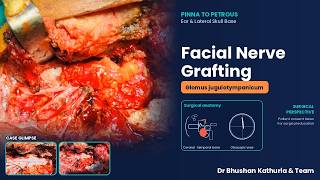 Chapter 13 · The Intratemporal Facial NerveFacial Nerve Grafting: Glomus jugulotympanicumLive surgery
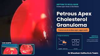 Chapter 16 · The Middle Fossa and Petrous ApexCholesterol Granuloma: Petrous apex, transnasal endoscopicLive surgery · petrous apex
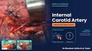 Chapter 17 · Completion, Complications and Transfer to the TheatreInternal Carotid Artery Control: Lateral skull base tumourLive surgery · from laboratory to theatre
Access and publication details
This edition is free to read online for personal educational use. Reproduction, adaptation or redistribution requires written permission from the author, except as permitted by law. The dissection record and score sheets may be copied for personal or supervised, non-commercial educational use with attribution. All other rights are reserved.
Self-published by Bhushan Kathuria, 1674P, Second Floor, Sector 2P, Rohtak 124001, Haryana, India. ISBN 978-93-345-8642-8 (electronic edition). The complete book can be downloaded as a PDF (52 MB) with chapter bookmarks. For corrections or permissions, contact kathuriabhushan56@gmail.com.
Back cover